Animals and medicines
The experimental protocol involving animals received full ethical approval from the Institutional Animal Care and Use Committee of Shanghai University of TCM (Approval No. PZSHUTCM2404280007). Male spontaneously hypertensive rats (SHR, 5 week-old) and age/sex-matched WKY rats were purchased from the Vital River Co., Ltd (Beijing, China). The rats were acclimatized in climate-controlled isolator cages and all procedures complied with humane animal testing guidelines. HQQR contains Salvia miltiorrhiza Bge., Ligusticum chuanxiong Hort., Uncaria rhynchophylla (Miq.) Miq. ex Havil., Haliotis diversicolor Reeve, Taxillus chinensis (DC.) Danser, Crataegus pinnatifida Bge. and Zea mays L. The detailed information on HQQR can be viewed in Supplementary Table 1 and the original drugs were purchased from Shanghai WanShiCheng Pharmaceutical Co. Ltd. Valsartan capsules were obtained from Novartis Pharmaceutical Co., Ltd and used as a positive control.
Reagents and antibodies
The primary antibodies included anti-β-myosin heavy chain (β-MHC, A22140, Abclonal, China), anti-atrial natriuretic peptide (ANP, A14755, Abclonal, China), anti-lactate dehydrogenase (LDHA, ab101562, Abcam, UK), anti-MPC1 (PS13040S, Abmart, China), anti-MCT4 (TD4182S, Abmart, China), anti-Mn-SOD (AB68155, Abcam, UK), anti-ATP6 (A8193, Abclonal, China), anti-p-PKM (#3827S, CST, USA), anti-glucose transporter-1 (GLUT1, A11727, Abclonal, China), anti-GLUT4 (PA3318S, Abmart, China), anti-hexokinase 2 (HK2, ab209847, abcam, UK), anti-GAPDH (60004-1-Ig, Proteintech, China) and anti-α-tubulin (66031-1-Ig, Proteintech, China). The fluorescent probe for rhodamine-labelled phalloidin (C2207S) was purchased from Beyotime (Shanghai, China). The angiotensin II (Ang-II, HY-13948) and VB124 (HY-139665) were purchased from MCE (USA). The pyruvate (A081-1-1) and lactate (A019-2-1) kit were from Jiancheng Institute (Nanjing, China). The lactate (L1750) was provided by Sigma (Saint Louis, MO). The rapid blocking buffer (PS108P) and ECL kit (SQ201) were from Yamei Biotechnology Co., Ltd (Shanghai, China).
Preparation of HQQR and identification of the main chemical components
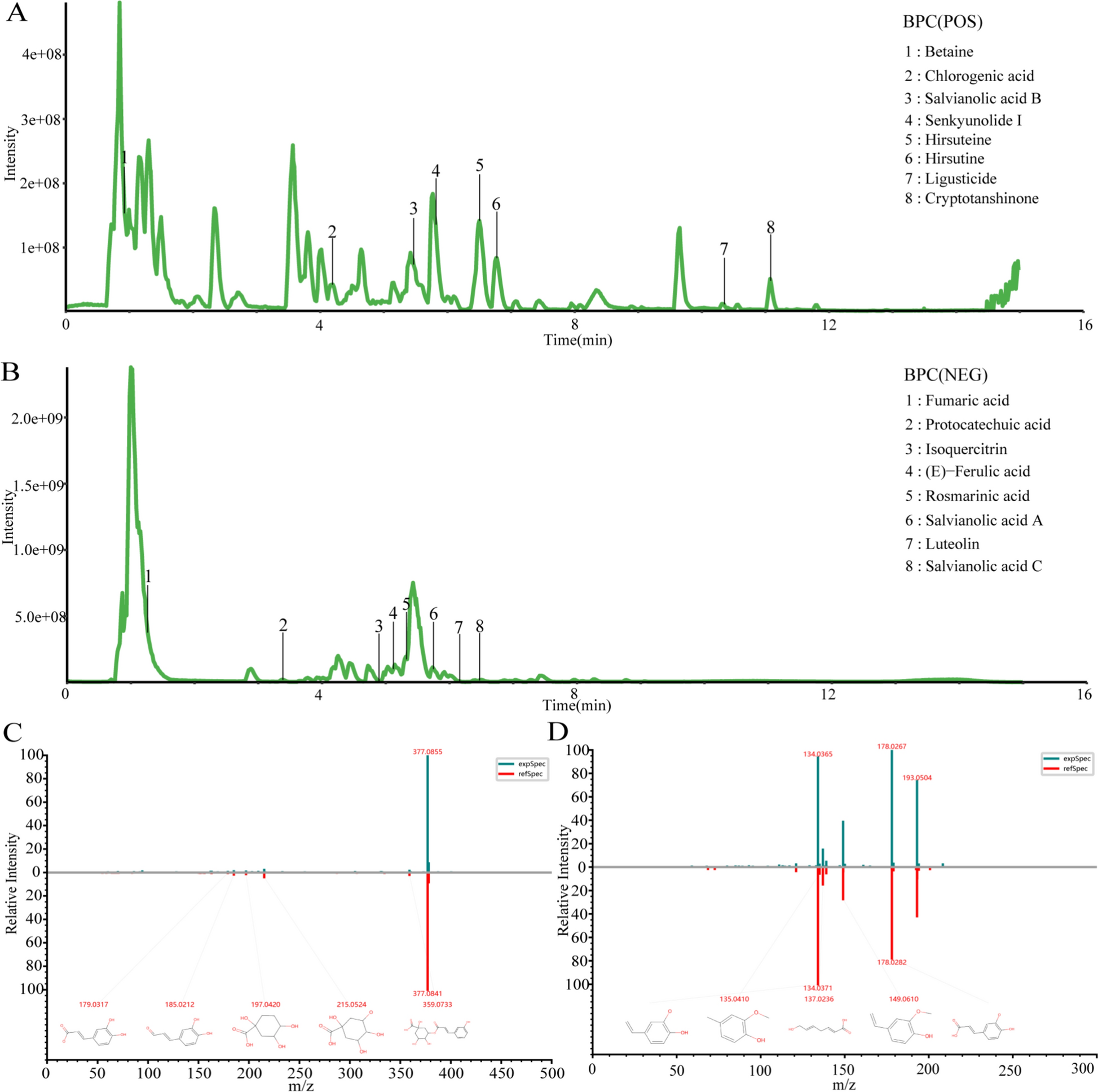
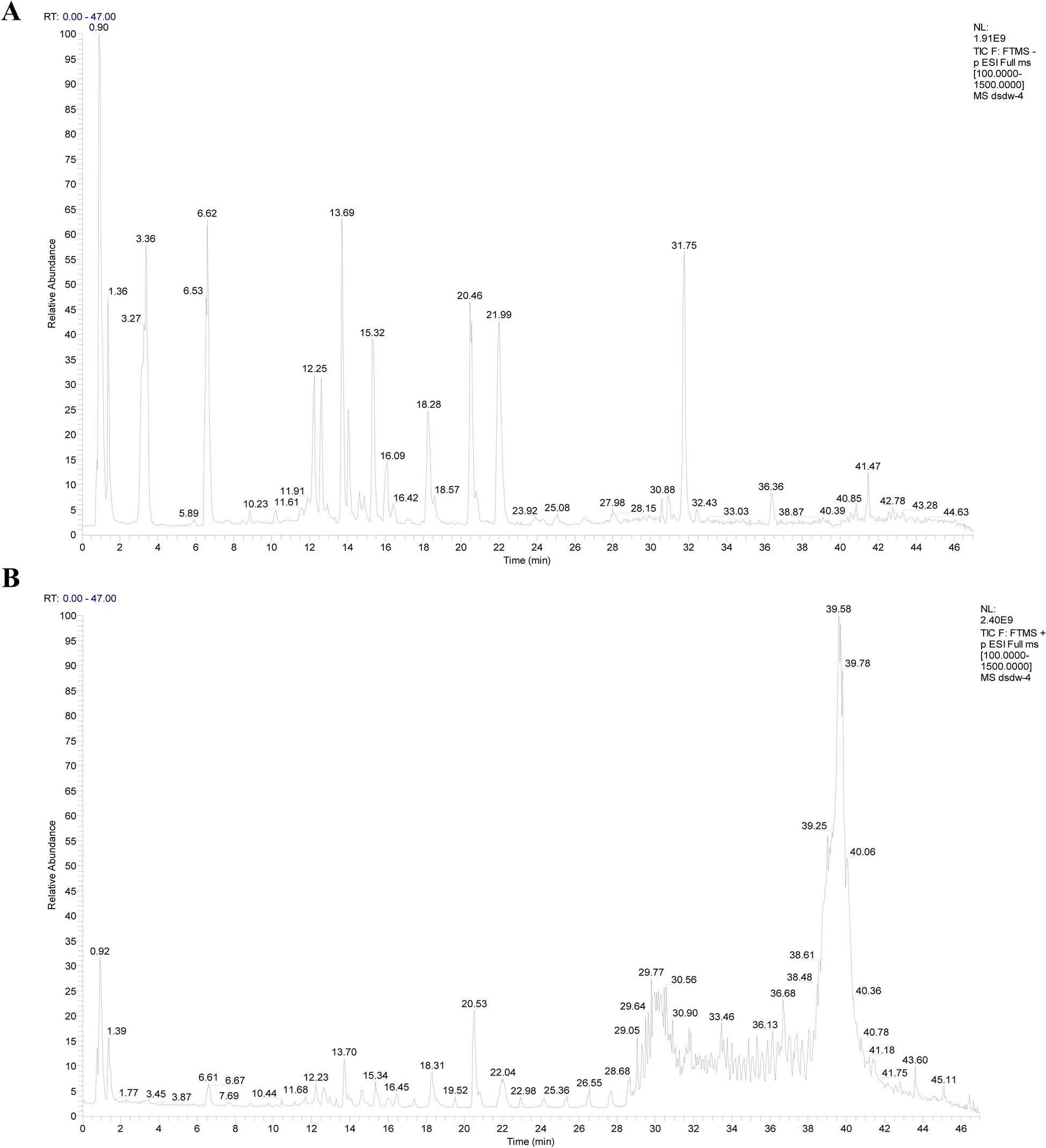
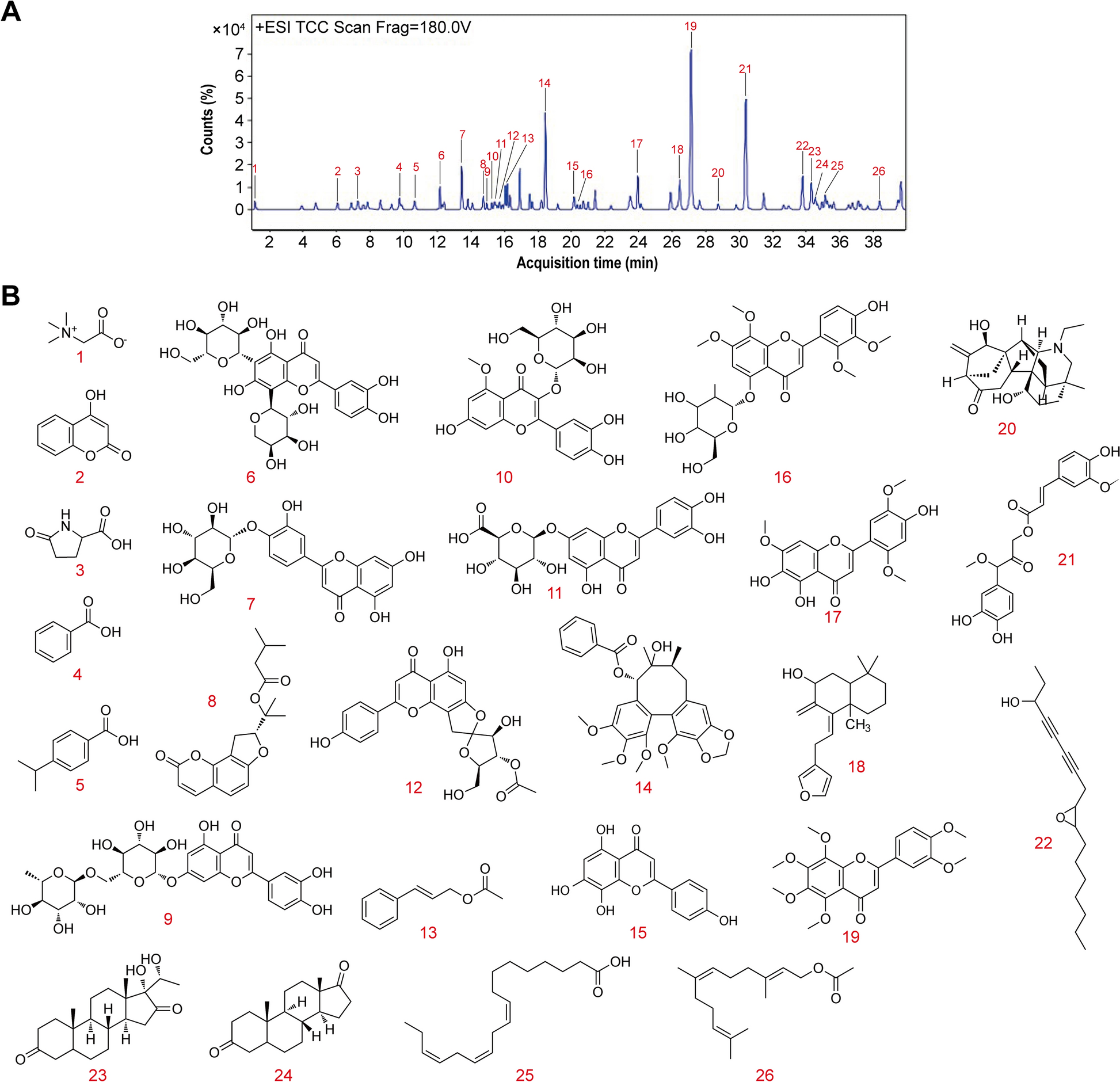
The aqueous extract of HQQR was prepared following our laboratory's optimized decoction protocol. The herbs contained in HQQR were mixed in proportion, crushed into small particles, and then boiled twice under continuous stirring. The aqueous extract was distilled and concentrated to approximately 3.87g (raw herbal product)/mL, and finally filtered. The chemical compositions of the aqueous extract were identified by Luming Biotech CO., Ltd. (Shanghai, China) using the ultra-high performance liquid chromatography-mass spectrometry (UPLC-MS) analysis. The column was an ACQUITY UPLC HSS T3 (100 mm × 2.1 mm, 1.8 μm) with a column temperature of 45 ℃. Mobile phase A was 0.1% formic acid–water, mobile phase B was acetonitrile, the flow rate was 0.35 mL/min, and the PDA scan range was 210–400 nm. The analysis was performed strictly according to the set elution gradient. According to the set mass spectrometry parameters, the mass spectrometry signal of the sample was collected using both positive ion and negative ion scanning modes.
HQQR-medicated serum preparation
Twenty male SD rats (220 ± 20 g) were selected for this study. These rats were then divided into two groups: an HQQR group and a control group, 10 rats in each group. Each rat was gavaged with 0.9% normal saline or HQQR solution (3.87 g/mL of drug) at a dose of 10 mL/kg twice a day for 3 consecutive days, respectively. Two hours after the final administration, rats were anaesthetised with sodium pentobarbital (40 mg/kg, i.p.). Blood was subsequently collected from the abdominal aorta and centrifuged to isolate serum. The serum was heat-inactivated in a 56 ℃ water bath for 30 min and sterilized by filtration through a 0.22 μm needle filter.
Animal model
The OBH rat model was prepared by feeding SHR rats with high-fat diet (HFD), and age/sex-matched WKY rats were used as controls. The high-fat feeds were obtained from Fanbo Co., Ltd. (Shanghai, China) and consisted of 60% normal feed, 4.7% raw peanut, 10% yolk powder, 1% sesame oil, 12% lard, 2% salt, 5% sucrose, 2% cholesterol, 3% milk powder, and 0.3% bile salt. After 10 weeks, rats in the top 1/3 of body weight were identified as OBH rats based on previous reports [32]. Next, the OBH rats were randomly divided into 4 groups (n = 8) and continued to be fed with HFD: (1) OBH-HF group, (2) OBH-HF/L group (5 ml/kg of HQQR, containing 3.87 g/ml of original drug), (3) OBH-HF/H group (10 ml/kg of HQQR), (4) OBH-HF/V group (10 ml/kg of Valsartan, containing 3 mg/ml of original drug). The other 8 SHR and WKY rats were divided into SHR-ND and WKY-ND groups, respectively, and fed normal diet. Each rat was gavaged once daily for 10 weeks.
Cell culture
The H9C2 cells, a rat cardiomyocyte, were purchased from the Pricella Co., Ltd (Wuhan, China) and cultured in DMEM supplemented with 10% FBS. To model myocardial hypertrophy, 85% confluence of H9C2 cells were treated with 1 μM of Ang-II and (or) free fatty acid (FFA) at 37 ℃ with 5% CO2 for 24 h. In this case, the cells were treated with or without HQQR-medicated serum. To observe the effect of lactate on cardiomyocytes, the H9C2 cells were treated with different concentrations of lactate (0.5 μM, 2 μM, 8 μM) with or without HQQR-medicated serum.
Western blot assay
Both myocardial tissues and H9C2 cells were lysed using RIPA buffer, and protein concentrations were quantified via the BCA assay. Proteins were separated electrophoretically on 8% SDS-PAGE gels and transferred to PVDF membranes. After sealing with a quick sealer, the PVDF membranes were incubated with primary antibodies according to the instructions. After incubation with secondary antibody to amplify the signal, the signal was visualised with ECL solution.
Pathology and oil red O staining
The 4 μm sections from the paraffin-embedded heart tissue were performed hematoxylin & eosin (HE) and Masson staining according to the standard protocol. For Oil Red O staining, OCT-embedded frozen sections were immersed in 60% isopropanol for 30 s and then incubated with Oil Red O staining solution for 15 min. Next, sections were placed in 60% isopropanol for colour separation, followed by a drop of Mayer's Hematoxylin staining solution for 2 min to stain the nuclei. Cardiac lipid droplets were observed under a light microscope at 200 × magnification and subjected to quantitative analysis using Image J software.
Wheat Germ Agglutinin (WGA) and phalloidin staining
Paraffin-embedded sections were sequentially deparaffinized in xylene and absolute ethanol, followed by antigen retrieval in EDTA buffer. Sections were incubated with WGA staining solution for 1 h at 37 ℃ protected from light, then counterstained with DAPI for nuclei visualization. For phalloidin staining, H9C2 cells were seeded evenly in six-well plates. After treatment, the medium was removed and cells were fixed with 4% paraformaldehyde. Following washes, cells were incubated with 1% phalloidin working solution for 30 min in the dark. Cardiomyocyte morphology was visualized using fluorescence microscopy at 200 × or 400 × magnification.
Determination of lactate/pyruvate ratio
The ratio of lactate to pyruvate was determined by the colourimetric method according to the instructions. Briefly, the serum was obtained by centrifuging the blood at 4 ℃ for 15 min. The serum was diluted in specific proportions and mixed with the test solution, then the mixture was reacted for 10 min at 37 ℃. Next, the mixture was incubated with the colour development solution for 10 min at room temperature. The absorbance values of lactate and pyruvate were measured at 530 nm and 505 nm wavelengths, respectively. The concentrations of lactate and pyruvate were calculated from the standard curves.
FFA determination
At the end of the intervention, blood was collected from the abdominal aorta of the rats and centrifuged to obtain serum. Serum FFA levels were tested by an automated biochemical analyser.
siRNA transfection
Rat MPC1 siRNA (5′-GCAAAGCAGCGGACUAUGUTT-3′) and MCT4 siRNA (5′-CUCAAUCGAUACUUCAACATT-3′) were synthesised by GenePharma (Shanghai, China). Negative control or target siRNA was transfected into H9C2 cells using lipofectamine 3000 reagent according to the manufacturer’s protocol. Briefly, cells were seeded in six-well plates to achieve 60–70% confluence. Lipid-siRNA complexes were prepared and added to cells for 12 h, after which the medium was replaced with fresh medium for subsequent treatments.
Statistical analysis
All experimental data were expressed as mean ± SD. Statistical significance was determined using ANOVA followed by LSD post hoc test for multiple comparisons, implemented in IBM SPSS Statistics 25.0 (IBM Corp., Armonk, NY, USA). A probability threshold of p ≤ 0.05 was applied to define statistical significance.


Comments (0)