
Remember me


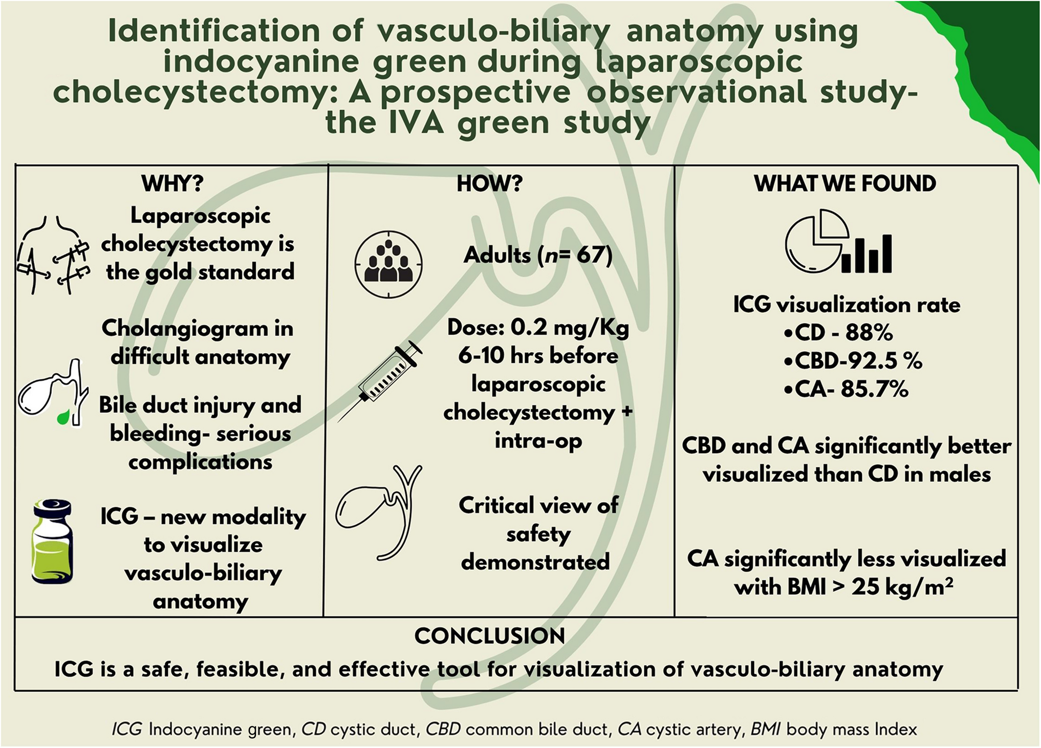
This study evaluated the feasibility and effectiveness of near-infrared fluorescence using indocyanine green (ICG) for intra-operative identification of cystic duct and artery anatomy, including aberrations, during laparoscopic cholecystectomy.
MethodsA prospective observational non-blinding study was conducted from November 2019 to February 2023, enrolling 67 adult patients scheduled for laparoscopic cholecystectomy. Indocyanine green (0.2 mg/kg) was administered six to 10 hours before surgery. Visualization of the cystic duct, common bile duct (CBD) and cystic artery was assessed using near-infrared fluorescence imaging. Univariate and multivariate analysis was done to identify the correlation between demographic and clinical factors with the identification of the cystic duct, CBD and cystic artery. Univariate and multivariate analysis was done to identify the correlation between gender and body mass index (BMI) with the identification of the cystic duct, CBD and cystic artery.
ResultsThe vasculo-biliary structures were visualized with ICG administration. The cystic duct was identified in 88%, the CBD in 92.6% and the cystic artery in 85% (Group-II variant was the commonest). Visualization of the cystic duct was significantly more difficult in male patients, while the visualization of the cystic artery was significantly reduced in patients with a > 25 kg/m2.
ConclusionsIndocyanine green-based near-infrared fluorescence is a safe, feasible and effective tool for enhancing the visualization of vasculo-biliary anatomy during laparoscopic cholecystectomy. It aids in preventing bile duct injury and supports safe dissection, particularly in patients with normal BMI. While not a replacement for intra-operative cholangiography, ICG fluorescence is a promising adjunct.
Graphical Abstract
Comments (0)