
Remember me


The investigators designed and implemented an uncontrolled clinical trial and the patients with OAF were recruited from the Oral and Maxillofacial Surgery Department Outpatient Clinic, Faculty of Dentistry, Tanta University, between September 2022 and May 2023.
The research for this study received approval from Tanta University’s Faculty of Dentistry Research Ethics Committee under code (#R-OS-9-22-6). Following the standards for human research approved by the Research Ethics Committee of the Faculty of Dentistry, Tanta University which follows the ethical guidelines outlined in the 1964 Helsinki Declaration and its subsequent revisions, the patients’ objective for participating in the study was described to them, and their informed consent was obtained prior starting treatment. NCT05987943 is the approved clinical trial number.
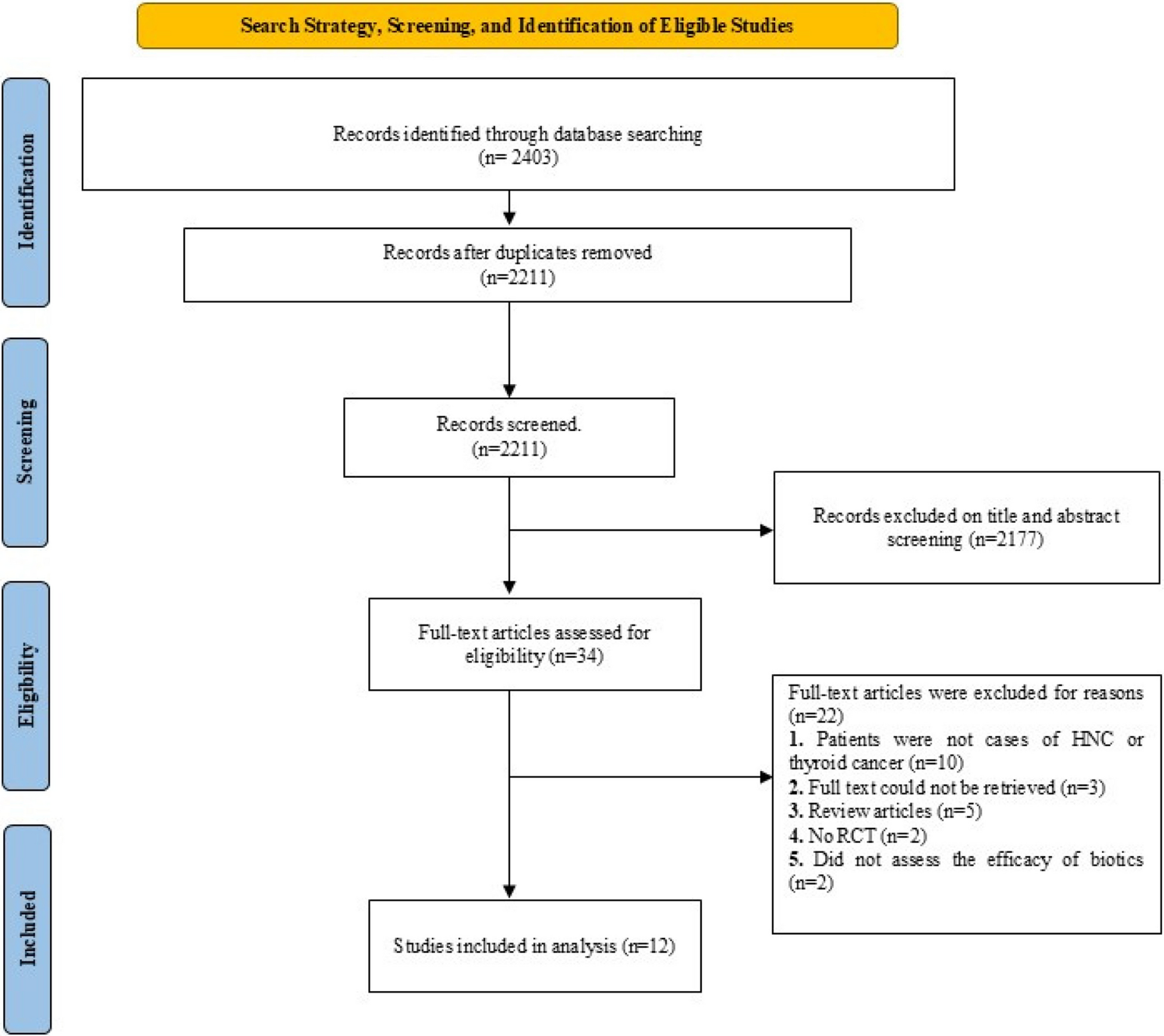
Eligibility criteriaPatients were selected according to the following main inclusion and exclusion criteria. The main inclusion criteria were patients complaining of nasal regurgitation of fluids, unilateral nasal discharge, changed nasal resonance, foul taste in the mouth, difficulty sucking via a straw and whistling sound when speaking and pain in the malar area. Patients with OAF less than 2 mm, debilitating diseases and not willing to participate in the study were excluded (Fig. 1).
Fig. 1 Sample size calculation
The sample size was calculated using G*Power software and sample size calculations Version 3.1.9.4. This power analysis used the difference in vestibular depth as the primary outcome. The effect sizes d = 1.33 was calculated based upon the results of Kumar et al. [11] and the estimated mean difference between the two groups = 0.18. Using alpha (α) level of (5%) and beta (β) level of (20%), that is, power = 80%; the minimum estimated sample size was a total of 7 subjects. The sample size will be increased to a total of 10 subjects to compensate for a dropout rate of about 20%.
Preoperative evaluation Chief complaintSymptoms (e.g., nasal regurgitation of fluids, unilateral nasal discharge changed nasal resonance, foul taste in the mouth, difficulty sucking via straw and whistling sound when speaking) are the main complaints. Pain in the malar area is possible.
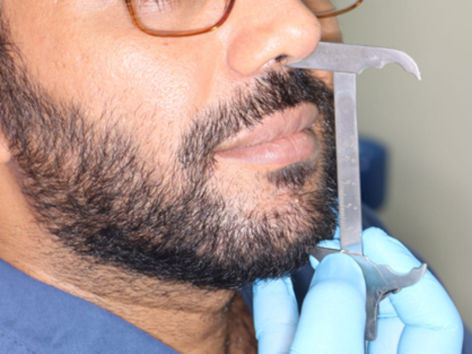
Clinical examinationValsalva test, cheek-blowing test, perforation investigation with probing and vestibular depth measured from gingival margin to bottom of the vestibule.
Radiographical examinationPanoramic radiograph to determine the location of the fistula as well as the existence and position of dental roots, implants, or any foreign bodies that may have become displaced within the maxillary sinus. Computed tomography (CT) scans were used to evaluate the magnitude of the underlying bone defect, exclude the occurrence of maxillary sinusitis, identify the existence of foreign bodies within the maxillary sinus cavity, and determine discontinuity of the maxillary sinus floor as shown in (Fig. 2).
Fig. 2
(A) Panoramic X-ray film demonstrating an oroantral fistula associated with the extracted upper left first molar. (B&C) A sagittal and coronal CT scan of the extraction site reveals an oroantral fistula, Case No. 2
Preoperative managementThe affected maxillary sinus should be irrigated with normal saline to eliminate infection through the fistulous orifice, followed by a betadine-containing solution diluted with normal saline. This process should be carried out until the lavage fluid is plain and without any inflammatory exudates.
Surgical procedureAll patients were treated with the pedicled buccal periosteal flap procedure under local anesthesia (4% articaine and epinephrine 1:100.000) using maxillary block and vestibular infiltration. To reveal the underlying connective tissue, a circular supra-periosteal incision was made with a 15-scalpel to eradicate the epithelial tissue along the fistula boundary. Following the elimination of the epithelial fistula wall, the sinus cavity was thoroughly curettaged and rinsed with saline solution to eliminate diseased and necrotic tissue.
A crestal incision was made with a Number 15 Bard Parker blade, with an anterior oblique releasing incision to the fistula. The buccal mucoperiosteal flap was split horizontally into two layers above the mucogingival junction: the first layer was a deep periosteal layer which was dissected submucosal about a distance of one tooth from the fistula while the second layer was a superficial buccal mucosal layer. Sutures were used to stabilize the pedicled deep periosteal layer, which was separated and turned above the oroantral fistula at the bone level and sutured to the palatal tissue. The superficial layer of buccal mucosa was bluntly dissected and sutured to the palatal tissue. In all cases, immediate evaluation with the Valsalva maneuver and probing with a blunt object revealed the main water-tight closure of the fistula as shown in (Fig. 3).
Fig. 3
(A) Preoperative photograph of an oroantral fistula (B) incision and reflection of buccal mucosal flap, (C) bony defect related to the oroantral fistula of upper left first molar, (D) reflection of periosteal flap from the underlying bone, (E) suturing of the periosteal flap over the oroantral fistula, (F) suturing of the buccal mucosa to the palatal tissue, Case No. 2
Postoperative managementPatients were advised to consume soft foods and avoid maneuvers such as sneezing with one’s mouth closed and nasal blowing that might raise the intrasinus pressure until healing occurred.
For two weeks, warm saline was used to keep the wound clean and the mouth was rinsed with 0.12% chlorhexidine digluconate mouthwash. To maintain antral orifice patent for drainage, all patients were given antibiotics, nonsteroidal anti-inflammatory drugs and nasal decongestants for at least 7 days.
Postoperative evaluationThe patients were observed weekly for one month, and later after three months. OAF closure was examined for healing, inflammation, infection, and any recurrence. Pain levels were measured using a 0–10 visual analogue scale (VAS), with 0 indicating no pain and 10 indicating the most severe pain, and the vestibular depth was measured from the gingival margin to the bottom of the vestibule using a periodontal probe.
Comments (0)