The rehabilitation of atrophic maxilla represents a challenge for oral and maxillofacial surgeon. Conventional implant treatments can’t be performed due to alveolar bone resorption and pneumatization of maxillary sinus. Since Bränemark discovered osseointegration [19], several surgical approaches for rehabilitating atrophic maxilla with dental implants have been proposed [20]. One of these approaches is maxillary sinus floor augmentation which is a predictable technique for obtaining the volumetric amount “vertical height” of bone required to place the implants [21, 22].
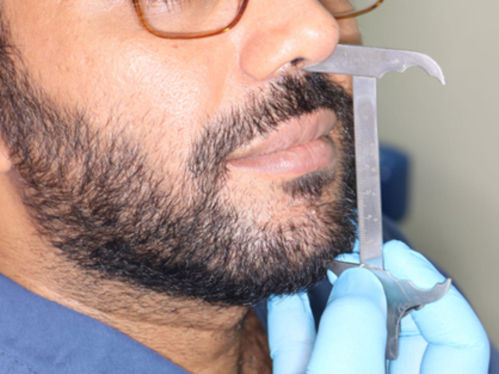
Sinus floor elevation can be executed via two distinct methods: the direct lateral window technique or the alternative indirect crestal method. The crestal approach to elevate the sinus floor is recognized for its minimally invasive nature, and its reduced complications when contrasted with the lateral window technique [23]. This method demonstrates enhanced predictability as a treatment option when the existing bone height is within the range of 5 to 6 mm, which is essential for ensuring primary stability [24].
The rationale of the present study was to compare the efficacy of Antral Membrane Balloon Elevation with Densah burs as minimally invasive techniques for atraumatic elevation of the sinus membrane through a transcrestal access.
The Antral Membrane Balloon Elevation (AMBE) technique is a minimally invasive approach, involves lifting the sinus membrane with minimal trauma, making it particularly advantageous for accessing challenging areas. Notably, it avoids sharp dissection around adjacent tooth roots, potentially reducing complications such as morbidity, blood loss, operative time, and postoperative pain compared to conventional methods [25]. The balloon technique can be applied to alveolar crests measuring 4 mm or less [8], in contrast to the classical indirect technique with osteotomes, where the minimum acceptable crest height is 6 mm [26].
A new surgical technique called osseodensification maintains bone by preparing the implant site by using Densah burs in anticlockwise direction (non-cutting motion) with copious irrigation. The osteotomy is gradually expanded in both the lateral and apical directions. The osseodensification concept is used for lateral expansion of narrow ridge, increase implant primary stability in low density bone and trasncresal sinus lifting procedure. The distinct flute design of Densah burs and counterclockwise rotation enable the lateral compaction of autogenous bone along the osteotomy walls and toward the sinus floor. This compaction, coupled with controlled pressure during drilling and a consistent in-and-out pumping motion, results in the creation of hydraulic pressure by well-hydrated autogenous bone, gently lifting and freeing the Schneiderian membrane [14, 15, 27, 28].
The choice of the bone grafting material to assist bone formation after sinus membrane elevation is still controversial [29]. None of the bone grafting materials on the market have all of the ideal properties as a bone substitute material [30, 31]. Autogenous bone grafting is widely recognized as the preferred method for grafting and reconstructive procedures in oral and maxillofacial surgery due to its established safety and reliability. However, it is also accompanied by several disadvantages, including the requirement for a second surgical site, heightened surgical risks, limited availability, and a relatively high rate of resorption [32].
In our study, allograft was used in both groups. Several studies have used allograft with maxillary sinus lifting procedures with simultaneous implant placement and had successful results [23, 24]. Allograft has osteoconductive properties, so it acts as a scaffold for bone formation [30, 33].
The survival rate in our study was 100% in both Groups. None of the cases showed any infection. Clinically and radiographically, all cases exhibited normal healing without any complications. Tang et al. [34] have reported that perforation of the Schneiderian membrane is the most common complication associated with transcrestal maxillary sinus lifting, occurring in 20% of cases. However, perforation was not reported in any case within the two groups, this could be attributed to the use of non-traumatic techniques, either by balloon or densah burs, which are capable of gentle slow elevation of sinus membrane.
Postoperative pain was examined using 10-point visual analogue scale (VAS) in the first 7 days. The two groups showed statistically significant decrease in pain score to reach score 0 by the end of the 7th day with no statistically significant difference between them. The pain experienced in both groups ranged from mild to moderate, consistent with findings reported by López-Quiles et al. [35].
Assessment of implant stability with the ISQ value of RFA is a non-invasive method widely used in the literature [36]. In this study, there was significant difference between the two groups in favor of Densah group at the time of implant placement (P = 0.004), but there was no significant difference between the two groups after 6 months (P = 0.07).
The notable disparity in primary stability between the two groups was largely attributed to the drilling protocol. In the Densah group, the spring back effect and elastic recoil of the bone on the implant surface following insertion resulted in an enhanced mechanical connection between the implant and the surrounding bone [37]. This finding aligns with the research of Huwais and Meyer [14], who noted that the osseodensification technique could lead to improved primary stability, a higher percentage of bone at the implant surface, and increased insertion and removal torques compared to the conventional drilling technique.
There was a significant difference when comparing implant stability immediately postoperative and at 6 months in the same group (P < 0.001). This is due to secondary stability establishment.
Our primary stability result was in line with Elghobashy et al. [37] and Hashem et al. [16], who compared the osseodensification technique with other techniques in crestal sinus lift procedures and found that the Densah group had the highest primary implant stability, but our result contradicts them regarding secondary stability.
Regarding residual bone height, it was almost the same in both groups, and there was no statistically significant difference between them (P = 0.624), indicating the homogeneity of the two groups at the start of the study.
In the present study, the mean immediate postoperative vertical bone height was 14.40 ± 0.53 mm in balloon group, while it was 12.70 ± 1.28 mm in Densah group, so there was a significant difference between the two groups in favor of balloon group (P < 0.0001) immediately postoperative, but there was no statistically significant difference between both groups after 6 months (P = 0.11).
Regarding the vertical bone gain in our study, the mean immediate postoperative vertical bone gain in balloon group and Densah group was 8.31 ± 0.44 mm and 6.75 ± 1.13 mm, respectively, so there was a significant difference between the two groups in favor of balloon group (P < 0.0001) immediately postoperative. After 6 months, there was no statistically significant difference between both groups (P = 0.25).
This is due to the fact that each 0.5 cc of the saline has an elevated sinus membrane of 6 mm in balloon technique [25], while Densah bur is used to pass the sinus floor and advance the height no more than 3 mm [38]. In addition, 0.5 cc of allograft was inserted in both groups which resulted in more elevation of the sinus membrane.
This was similar to López-Quiles et al. [35], and Abdalhameed et al. [25] who had 8.22 mm and 7.27 mm of immediate postoperative vertical bone gain, respectively after a crestal sinus lift procedure using antral membrane balloon elevation. Also, the result of our study was in line with Hashem et al. [16], who had 5.81 mm of vertical bone gain after crestal sinus floor elevation using Densah burs.
In contrast to our study, Ismaeil et al. [39], reported 4.55 mm of vertical bone gain after crestal sinus lift using balloon technique after 3 months of implant insertion.
The IPL appears to be a critical factor affecting new bone formation. Our study demonstrated that the mean IPL was 3.66 ± 0.88 mm in balloon group and 3.80 ± 1.07 mm in Densah group, with no statistically significant difference between the two groups (P = 0.689). When IPL is between 3 and 5 mm, new bone formation will be of a great amount. Our findings are consistent with those of Lin et al. [40].
Regarding reduction in vertical height, the mean was 2.28 ± 0.57 mm and 1.13 ± 0.40 mm in balloon group and Densah group, respectively, with statistically significant higher vertical height reduction in balloon group than Densah group (P < 0.0001). This could be attributed to the standardization of the quantity of bone graft for each implant at 0.5 cc allograft. In addition, the greater the elevation of the sinus membrane in balloon group, the greater the membrane tension will be, and the tension may be transferred to the force of compression on the grafting materials [41, 42].
Similar to our results, Jensen et al. [43], found that the resorption rate of bone is impacted by the types of graft materials. They observed resorption rates of 1.8 mm in autograft, 2.1 mm in demineralized allograft, 0.9 mm in alloplast, and 0.8 mm in autograft mixed with alloplast.
Regarding the bone density, there was no significant difference between the two groups in preoperative buccal and palatal bone density (P = 0.256, P = 0.587), respectively, which indicated the homogeneity of the two groups at the start of the study. And, throughout the study, the buccal and palatal bone density showed a significant increase from baseline to 6 months in the same group. This increase in bone density was related to the technique itself.
There was a significant difference in the buccal and palatal bone density between the two groups in favor of Densah group immediately postoperative and after 6 months. This is due to osseodensification technique using Densah burs cause motorized expansion of osteotomy site which results in increase the bone density around the implant. This is in line with other studies of Kumar et al. [44], and Huwais and Meyer [14]. This increase in bone density in Densah group is coincide with the high primary stability, where the stability depends on close contact between the implant surface and the surrounding bone [14].
The limitations of this study are a small sample size and a relatively short follow up period. An extended follow up period may yield a more pronounced result. So, greater sample sizes and longer follow-up periods should be the main goals of future research.


Comments (0)