The VDO may be described as the superior limit of rotation closure of the mandible around the bicondylar hinge axis. Hence, with the loss of posterior teeth, there will be a loss of VDO [19, 31].
The routine clinical determination of the resting position for the measurement of lower facial height is not accurate for establishing the appropriate lower facial height. The remaining position is not fixed, as it could be influenced by a variety of extrinsic and intrinsic factors [32].
There are many other methods available for estimating the VDO. However, these methods are empirical in nature, and currently, there is minimal scientific proof or a universally accepted method for the precise determination of VDO. To date, there has been no method for precisely measuring VDO. The traditional method relies on a subjective adjustment (subtracting 2–4 mm from VDR), which can vary based on the clinician’s experience and judgment. This study used the distance between the mental foramina, which provides an objective and consistent anatomical landmark visible on panoramic radiography. This reduces the variability and subjectivity associated with the traditional method. By providing a precise and objective measurement, our method reduced the risk of errors in VDO estimation. Incorrect VDO can lead to complications. Anatomical and radiographic studies ensure that VDO is a more accurate compared to the empirical subtraction method, which may not account for individual anatomical variations. [8, 11, 15, 19, 20, 25, 33] Hence, it was necessary to identify a method that can guide the prediction of lost facial dimensions.
Panoramic radiographs were selected because they are routine dental diagnostic aids and are usually included in patient records. Panoramic radiography provides a detailed view of the maxillomandibular area, presenting a unique image of both the upper and lower dental arches. This imaging method provides a better view of the bone structure, especially the lower jaw (mandible), and could be a good guide for examination prior to prosthetic rehabilitation. Other advantages of this technique include a lower radiation intake for the patient and a relatively shorter imaging time [34].
To achieve high reliability of the collected data, examiners were trained through a series of clinical trainings and calibrated using the protocol of this study to avoid any bias. Kappa coefficients for categorical data were used to explore the reliability of the data [35]. The kappa coefficient is considered the statistic of choice to analyse the reliability of nominal and categorical types of data recorded on the same patient by more than one clinician. [36] Inter- and intraexaminer reliability were calculated using kappa statistics and were found to be 0.87 and 0.78, respectively. The calculated root of the mean squared error (RMSE) between the actual and estimated VDO was 0.133 cm.
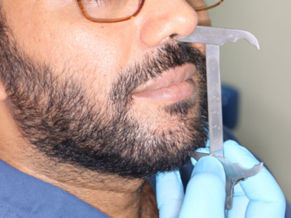
VDO was measured using two easily recognized and stable points, the columellar base septum of the nose and the base of the chin. This study proposed a novel formula between the VDO and the intermental foramen distance (IMFD) using linear regression analysis between the VDO and IMFD. The proposed formula was (VDO = 0.98 × IMFD + 0.45 cm).
After generating the formula, it was used with an additional 50 subjects and yielded promising results. The difference margin between the actual VDO and the formula is -0.02 to + 0.25 cm. This results was consistent with the results from the Bland‒Altman plot test between VDO and IMFD, which is a test used for an alternative analysis based on the quantification of the agreement between two quantitative measurements by studying the mean difference and constructing limits of agreement [37].
The mean VDO in this study was 6.8 ± 0.52 cm. These results were in agreement with the results of Miran [38], who studied the role of anthropometric measurements in determining the occlusal vertical dimension and found that the mean OVD in the Kurdish population was 6.863 cm in males and 6.199 cm in females.
Preetha et al. [39] conducted a study in the Indian population to assess the relationship between the length of the right ear, occlusal vertical dimension, and length of the right index and little finger; they found that the mean occlusal vertical dimension was 7.142 ± 0.483 cm.
Helal [40] evaluated lower facial heights as related to different anthropometric measurements in dentate and completely edentulous subjects and reported that the mean lower facial height from the base of the chin to the subnasal point was 6.46 ± 0.81 cm. This difference may be attributed to the use of different patient samples, as he conducted his study on male Arabian patients in Saudi Arabia.
The mental nerve is a key factor in many surgical and clinical procedures in routine clinical practice [41]. The mental foramen was chosen because it is a landmark that can be easily interpreted on panoramic radiographs due to its fixed, known location and obvious radiographic features of radiolucent rings with sharply demarcated radio-opaque borders on radiographs throughout life, including in older populations. Extensive literature indicates that the visibility of the mental foramina on panoramic radiographs does not significantly diminish with age. Therefore, the reliability of these landmarks as reference points for measuring the vertical dimension of occlusion (VDO) remains consistent across different age groups [42, 43].
The mean intermental foramina distance [IMFD] from the distal border of the mental foramen to the same point on the other side was 6.47 ± 0.51 cm. In males, it was 6.62 ± 0.47 cm, whereas in females, the mean IMFD was 6.2 ± 0.50 cm.
Nejaim et al. [44] utilized panoramic radiographs to compare the distance and symmetry of the mental foramen observed in situ with those in radiographic images in a Brazilian sample. They studied 58 mandibles both in the laboratory and radiographically using digital panoramic radiographs. The distance from the mesial aspect of the mental foramen on the right side to the mesial aspect of the mental foramen on the left was found to be 4.481 ± 0.176 cm. This differs from the mean distance in our study (6.47 ± 0.51 cm). This discrepancy can be explained by several factors: the panoramic radiographs in our study were taken from living subjects, whereas Nejaim et al.‘s study involved macerated mandibles from a different population. Additionally, our measurements were taken from the distal aspect of the mental foramen, while Nejaim et al. measured from the mesial aspect.
Assessment of mental foramen position relative to anatomical landmarks was done by Sheikhi et al. [45] for 180 CBCT radiographs for Iranian population. The mean distances from the anterior border of the mental foramen to the midline were 2.586 cm (SD ± 0.027) on the right side and 2.553 cm (SD ± 0.031) on the left side. Summing these distances gives a total of 5.139 cm between the mesial borders of the right and left mental foramina. By adding the average diameter of the mental foramen to their findings to standardize the measurement method with our study, the distance becomes 5.857 cm. This is 0.613 cm different from our study’s findings. This discrepancy can be attributed to the different radiographic methods used: Sheikhi et al. employed CBCT, whereas our study utilized digital panoramic radiographs.
Similar results using CBCT were found in a study conducted by Kabak et al. [46]. The average distance between the right and left mesial surfaces of the mental foramina, according to the data of their study, was 5.05 cm.
In agreement with our study, Mohamed et al. [47]. compared the location of the mental foramen across different age groups using digital panoramic radiography in 250 subjects. They measured the distance between the centres of the mental foramina from the right to the left side, finding an average distance of 6.234 cm. This measurement closely aligns with our study, which found an inter-mental foramen distance (IMFD) of 6.47 cm. The slight difference is due to our measurement being taken from the distal aspect of the mental foramen rather than from the centre.
This study facilitated the determination of the lower facial height, aiding in the construction of full or partial prostheses and those patients with broad prosthetic rehabilitation. The formula obtained in this study could be further studied by applying it to edentulous patients to determine the clinical applicability of the derived formulae. Additionally, software or an application could be created to calculate the dimensions directly, which can also be an innovative trend.
Limitations
The limitations of this study include that it was conducted only for subjects with class I malocclusion, and other dental and skeletal malocclusions were not included.
The fact that the study sample was limited to Egyptian subjects is another limitation. The relevance of the current findings to other ethnic groups must thus be assessed through additional research using the same methods in diverse populations.
This study was performed on relatively young individuals in which the effect of ageing was not observed. Therefore, further studies should examine elderly subjects among whom chronological changes are likely to occur.


Comments (0)