
Remember me


The trial was conducted as a randomized clinical study with two parallel groups that had the same allocation ratio and was planned as a double-blinded study including assessors and patients. ClinicalTrials.gov formally registered the trial protocol with the ID number (NCT05970536). The trial was conducted from December 2022 to December 2023 in the outpatient clinic at the faculty of Dentistry Kantara branch- Sinai University. In order to ensure clear and transparent reporting in this research, the CONSORT 2010 criteria (Consolidated Standards of Reporting Trial) were followed. Additionally, the Faculty of Dentistry and Cairo University’s Research Ethics Committee approved this trial (34/11/22). Before the study began, written informed consent forms were signed by each participant. For the volunteers’ ease of understanding, All consent form was created in Arabic. Participants were given a clear explanation of the trial’s objectives, benefits, risks, and anticipated duration in plain language.
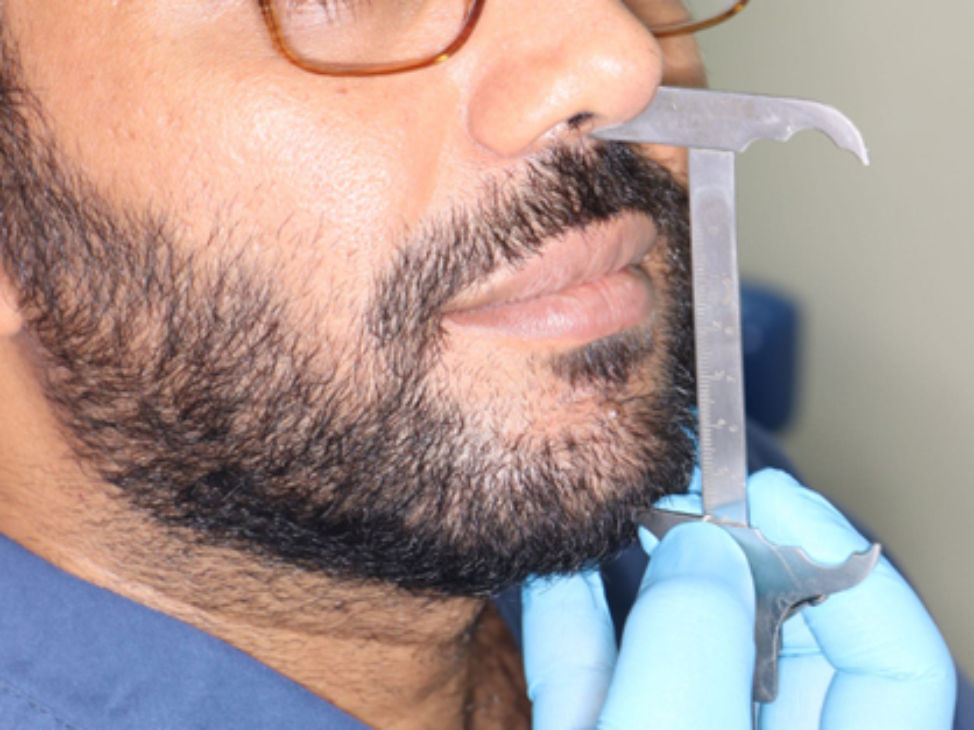
Eligibility criteriaForty patients in the 18–50 age range who were in systemically good health were screened for trial enrollment. The chosen patients received training in dental hygiene along with a full mouth scaling procedure. Considering the study’s inclusion and exclusion criteria, a single session of treatment was conducted for each patient exhibited an exceedingly deep carious pulp exposure with a mature closed apex and normal apical tissue.
Following the completion of the history, clinical and radiographic examinations, as well as cold and electric pulp tests, were conducted to test for pulp sensibility; consequently, the diagnosis of periapical and pulpal tissue was established. Based on the history of persistent or unplanned pain was replicated by cold trials, a clinical diagnosis consistent with symptomatic irreversible pulpitis (SIP) was made (Endo-frost, Coltene, Whaledent GmbH). The results of inclusion showed that the teeth had a healthy periodontium, no radiographic evidence of periapical radiolucency, and no unfavorable reactions to percussion or palpation testing.
Exclusion criteria included patients taking analgesics during the last week, taking antibiotics within the last month, experiencing insufficient bleeding or being unable to control bleeding within five minutes of pulp exposure, pregnant females, or having medical issues and history of intolerance to anti-inflammatory drugs. While the teeth exclusion criteria were sinuses, swelling, a negative response to vitality test, non-restorable and had periodontal involvement or mobility, lacked pulp exposure or underdeveloped roots even after excavation of caries that had previously undergone treatment. Depending on clinical findings, patients who met the eligibility requirements were given information on VPT as well as the procedure’s risks and benefits. After that, signed informed consent was then acquired.
Sample size calculationMedCalc® version 12.3.0.0 program “Ostend, Belgium” was used for calculations of sample size, statistical calculator based on 95% confidence interval and power of the study 80% with α error 5%, According to a previous study [18], showed that the percentage of perceptible grey discoloration in ProRoot MTA (80%) compared to Biodentine group (26.9%). So it can be relied upon in this study, based on this assumption, sample size was calculated according to these values produced a minimal samples size 28 sample were enough to find such a difference. So, by calculation, the sample size will be equal to 14 sample per group.
RandomizationThe entire pulpotomy will be finished up to the point where hemostasis can be easily obtained after pulp exposure has been clinically established. Subsequently, participants were randomized to the EDTA or apple vinegar groups. Using a Microsoft ® Excel program, a computer-generated randomization was used to randomly allocate participants to each of the assigned treatments in the clinical trial. A list of sequential numbers was created, in which each randomly assigned participant in this list occupied a sequence number (ID) from “1 to 40” and was also given another randomized number represents either group 1 where the final irrigating solutions was 17% EDTA or group 2 where the final irrigation was apple vinegar. The generation of random allocation was done by Dr. (H R) who was not involved in this clinical trial and was independent from the recruitment process. The allocation sequence of the assigned participants in this clinical trial was kept with (H.R) in a sealed envelope which was known at the time the participant was given his informed consent to be involved in the clinical trial through contacting (H.R) by phone. The operator was not blind in to the study.
Clinical procedureEvery patient had a pulpotomy procedure carried out by one operator utilizing aseptic measures and customary procedures. To achieve deep anesthesia, 2% lidocaine and 1:100 000 epinephrine (Ramson Remedies) was applied. Then, the tooth was sealed off with a rubber dam and cleaned with cotton pellets dipped in 5.25% sodium hypochlorite (NaOCl; Ammdent). Caries was removed with a sterilized big round diamond bur (ISO 001, Mani) and spoon excavators with high-speed hand pieces submerged in water coolant. A tapered round diamond bur was used to remove any residual supporting tooth structure. Composite resin (Fusion light-cured universal nanohybrid composite, Prevest DenPro), and sectional matrix (Saddle contoured metal matrices, Filaydent) were used to reconstruct missing proximal surfaces.
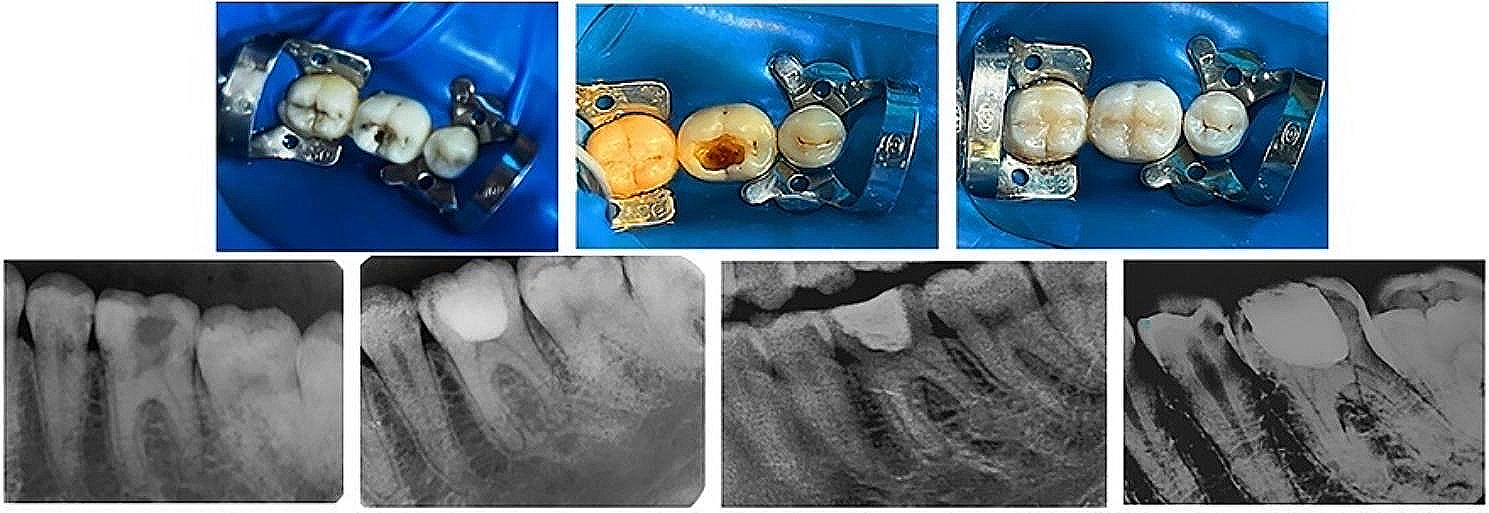
An aseptic tapered diamond bur was employed to refine the opening following pulp exposure. The vital pulp was clinically determined by the presence of pulpal bleeding. utilizing a water-cooled, high-speed hand piece with a sterile big round diamond bur, the exposed tissue of coronal pulp was gently removed Then, it was severed at the point where the root canal openings until hemostasis could be reached Fig. 1 Cotton pellets moistened in 3% NaOCl (Parcan, Septodont) were applied to the pulpal wound for a duration of two minutes, and up to five minutes if necessary till hemostasis occured.
Fig. 1
Clinical Photograph showed: Preoperative, full pulpotomy with hemorrhage control, Composite restoration. Clinical Radiographs showed: Preoperative, Postoperative at 3, 6 and 12 months, respectively
RCT was used to handle all cases, regardless of the assigned group, where hemorrhage could not be halted within five minutes. Before the last treatment, the field was first cleansed with 5 ml of 1.5% NaOCl and then submerged in saline irrigation. Following hemostasis, the chosen teeth were randomly separated into two primary groups based on the final irrigating solutions: Group 1: A 17% EDTA for five minutes was used on exposed pulp tissue. Group 2: Apple vinegar was used to irrigate exposed pulp tissue for a duration of five minutes. According to the manufacturer’s recommendations, a bioceramic material ProRoot white MTA (Dentsply, DeTrey GmbH) was just mixed in both groups, transported to the pulpal wound utilizing an amalgam carrier, and gradually thinned to a thickness of 2–3 mm with dampened cotton granules to facilitate MTA settling, followed by a layer of glass ionomer as a temporary restoration for the coronal seal for some participants who have no time to stay for permanent composite restorations (GI; KetacTM Molar, 3 M Deutschland GmbH).
Consequently, the following day for those participants, the rubber dam was applied the cotton pellet and temporary restoration were withdrawn, and the MTA setting was verified. Afterwards MTA was covered with a thick coating of glass ionomer (KetacTM Molar, 3 M Deutschland GmbH) and composite resin as permanent restoration while for other participants permanent composite restorations were applied immediately over glass ionomer after vital pulp therapy procedures.
(Fusion light-cured universal nanohybrid composite, Prevest DenPro) was applied, polished, completed and corrected for occlusal contacts for all the included participants A Carestream RVG 5200 digital imaging system was used to take an instantaneous postoperative intraoral periapical radiograph (Carestream Health Inc.).
OutcomesThe primary outcome revealed that the patients were returned back after three, six, and twelve months to evaluate the results. As recommended [19], the outcome assessment included both a clinical examination, a pain assessment and apulp response to a thermal and electrical pulp tester. Clinical evaluations were performed on teeth to determine whether any periapical or pulpal diseases were present, as well as their signs and symptoms. The pain assessment included patients that were told to utilize the 0–3 Visual Analogue Scale (VAS) to score their pain level [20]. Preoperative pain was documented by the patient at baseline, and postoperative pain was documented on VAS scores spaced out by 2, 6, 24, 48, and 72 h. There were four categories for pain scores: mild (Vas score 1), moderate (Vas score 2), severe (Vas score 3), and no pain (Vas score 0). The participants were instructed to go back to the clinician if their acute pain persisted even after taking the prescribed medication, and they were given instructions to take analgesics (Ibuprofen 400 mg every 6–8 h) for pain alleviation if necessary. Since ibuprofen has dose-dependent activity and its analgesic effect completely disappears after 8 h, these patients were evaluated at 24, 48 and 72 h similar to other patients in the study.
The secondary outcome showed the radiographic criteria which indicated absence of pathosis or resorption after three, six, and twelve months to evaluate the results. Lack of clinical and radiographic manifestations denoted that the treatment was deemed successful.
For statistical analysis, the statistical software for social sciences, version 23.0 (SPSS Inc., Chicago, Illinois, USA), had been used to evaluate the recorded data. An analysis of significance using paired samples t-test was performed when comparing related samples. ANOVA tests with repeated measures were used to see if there were any differences between related means. Bonferroni correction was applied in post-hock comparisons. To compare two means, the independent-samples t-test of significance was employed. Numbers and percentages were utilized to represent the qualitative characteristics. The Chi-square test and Fisher’s exact test were applied to compare groups with qualitative data. The likelihood, P-value was reported: P-value > 0.05 was deemed inconsequential, P-value < 0.05 was deemed noteworthy, and P-value < 0.001 was deemed extremely significant.
Comments (0)