
Remember me


A 16-year-old male adolescent visited the Hospital of Stomatology, Xi’an Jiaotong University, with the chief complaint to continue his orthodontic treatment and address severe tooth surface demineralization. Diagnosed with a unilateral complete cleft lip on the right side at birth, he underwent cleft lip repair and received speech therapy in his infancy and early childhood. The patient has no relevant family history. At the age of nine, the patient underwent alveolar bone grafting surgery. Approximately two years prior to this visit, he began orthodontic treatment for permanent teeth at another dental clinic. However, the treatment was halted due to persistent spaces in the upper arch, severely misaligned occlusion, and extensive tooth demineralization/caries, which the previous dentist was unable to manage.
The patient exhibited a scar extending from the right upper lip to the base of the nose, resulting in asymmetrical peaks of the upper lip. Additionally, nasal asymmetry was evident, with a collapsed right nostril. His facial profile revealed protrusion of both the upper and lower lips (Fig. 1a). Fixed orthodontic appliances were bonded to the surfaces of teeth, accompanied by a removable occlusal splint for the maxillary posterior teeth. Moreover, the adolescent’s oral hygiene was notably poor, characterized by significant accumulation of calculus and plaque around the brackets, even on the front teeth (Fig. 1b). Upon examination with a dental probe, the surrounding areas of the brackets felt soft. After the occlusal splint was removed, the patient presented bilateral Class II relationships for both canines and molars, with an overjet of 4 mm and a pronounced deep overbite (Fig. 1c). A fissure was observed in the gingiva distal to the right upper central incisor, corresponding to the location of the alveolar cleft (Fig. 1b). The right upper lateral incisor displayed microdontia, accompanied by a 6 mm space mesial to this tooth (Fig. 1b). The bilateral mandibular posterior teeth were lingually inclined, resulting in a scissor bite of the right premolars and first molars, while the left premolars and molars showed a significant deep overjet (Fig. 1b and c).
Fig. 1
Pre-retreatment examinations of the adolescent with unilateral complete cleft lip. (a) Pretreatment facial photographs. (b) Pretreatment intraoral photographs. Notably, the patient showed considerably poor oral hygiene, characterized by significant accumulation of calculus and plaque around the fixed brackets. (c) Pretreatment dental casts without a removable occlusal splint for the maxillary posterior teeth. The rear view displays significant discrepancies in the width of posterior teeth, including a scissor bite of the right premolars and first molars and a deep overjet of the left premolars and molars
The pretreatment lateral cephalometric assessment revealed a skeletal Class I relationship (ANB = 2.2°) and a low mandibular plane (GoGn-SN = 21.9°). The maxillary incisors were slightly forward-leaning (U1-NA = 7.7 mm), whereas the mandibular incisors were slightly lingually inclined (L1-NB = 3.1 mm) (Fig. 2a and Table 1). The pretreatment panoramic radiograph showed three impacted third molars and a shortened root for the right upper lateral incisor (Fig. 2b). In addition, the pretreatment CBCT indicated the presence of bone at the site of the alveolar cleft bone graft (Fig. 2c), along with a considerable amount of bone on the buccal sides of the bilateral mandibular molars, which were lingually inclined (Fig. 2d).
Fig. 2
Pre-retreatment lateral cephalogram, panoramic radiograph and CBCT. (a) Lateral cephalogram. (b) Panoramic radiograph. (c-d) CBCT. An axial slice revealing bone presence at the alveolar cleft bone graft site, located distally to the right maxillary central incisor (indicated in c), and a coronal slice at the level of the second molars showing significant discrepancies in the width of posterior teeth bilaterally (indicated in d)
Table 1 Cephalometric measurementsBased on the medical history and examinations, the diagnosis for the adolescent included UCCL, a bone-grafted alveolar cleft, and enamel demineralization resulting from orthodontic treatment.
Treatment objectives and strategyThe treatment objectives were to: (1) improve the patient’s oral hygiene and address the demineralization of enamel, (2) establish an ideal functional occlusion for both anterior and posterior teeth, and (3) achieve dental and facial aesthetics.
The treatment strategy included: (1) removal of the existing fixed appliances, remineralization of the teeth, and providing instructions for oral hygiene, (2) aligning the upper teeth and reducing the overbite with a flat anterior bite plate, (3) uprighting the lower molars that lingually inclined, (4) closure of the space in the upper arch and preservation of space for the prosthetic management of the microdontia, (5) surgical interventions to enhance nasolabial esthetics, and (6) use of a Hawley retainer equipped with a flat anterior bite plate to ensure the long-term stability of the treatment outcomes.
Treatment methodology and progressInitially, we removed all orthodontic appliances and conducted supragingival scaling, followed by the remineralization treatment for this adolescent. For remineralization therapy, a fluoride varnish containing 5% sodium fluoride was utilized (3M™ Clinpro™ White Varnish). Fluoride varnish application was performed after thorough plaque removal and complete drying of the tooth surfaces. A thin layer of varnish was evenly applied using a small brush. The treatment was repeated after four weeks, and the patient was instructed to follow up every three months to assess the need for further in-office fluoride varnish treatments. Additionally, the patient was instructed to use toothpaste containing 5,000 ppm sodium fluoride (Colgate® PreviDent® 5000) twice daily. Crucially, the patient was provided with comprehensive and rigorous oral hygiene education to improve his dental care practices. After two months, a notable improvement in oral hygiene was observed, along with effective remineralization of the enamel (Fig. 3a). Quantitative assessments [13, 14] indicated a notable decrease in both the Oral Hygiene Index-Simplified and the Plaque Index (Table 2), confirming substantial oral hygiene improvement.
Fig. 3
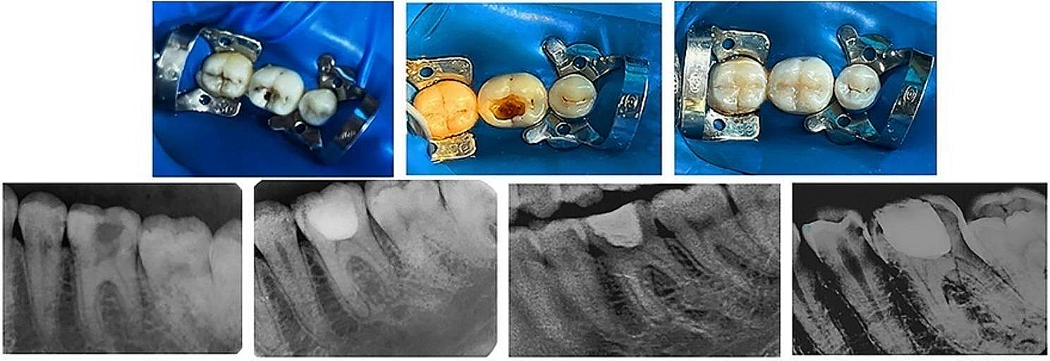
Progress intraoral photos during the treatment. (a) The remineralization treatment was completed, and a fixed straight wire appliance (SWA) was bonded to the upper teeth, accompanied by the placement of 0.012-inch nickel-titanium (NiTi) archwire. Notably, compared to the pre-retreatment condition, a notable improvement in oral hygiene was observed, along with effective remineralization of the enamel. (b) 5-month stage of the orthodontic treatment. A removable flat anterior bite plate was employed to address the deep overbite, and brackets were bonded to the lower teeth. (c) 11-month stage of the orthodontic treatment. Lingual segmental arches were applied on the lower molars (indicated by arrowheads), and additional labial crown torque was employed to the main archwire to correct lower molars that lingually inclined. (d) 14.5-month stage of the orthodontic treatment. The deep anterior overbite was corrected, and the lower molars were uprighted. Space closure was begun using sliding mechanics. (e) 26.5-month stage of the orthodontic treatment. The fixed orthodontic appliance was removed. At this stage, aesthetic prosthodontic treatment for the microdontia of the right maxillary lateral incisor had not yet been performed
Table 2 Quantitative assessments of oral hygieneSubsequently, fixed straight wire appliances (SWA, Shinye lnc., China) were bonded to the maxillary teeth, and a 0.012-inch nickel-titanium (NiTi) archwire was placed (Fig. 3a). The alignment and leveling of the upper arch were achieved through the sequential replacement of archwires, and a removable flat anterior bite plate was employed to reduce the deep overbite of the patient (Fig. 3b). By the 11th month of the orthodontic treatment, after placement of 0.018 × 0.025-inch stainless steel (SS) archwires on both arches, lingual segmental arches on the mandibular molars and additional labial crown torque on the main mandibular archwire were utilized, to correct lower molars that lingually inclined (Fig. 3c). At the 14.5-month juncture of the orthodontic treatment, with the anterior deep overbite corrected and the mandibular molars uprighted, space closure was begun using sliding mechanics (Fig. 3d). Class II elastics (1/4, 3.5 oz) were utilized to achieve Class I relationships for the canines and molars. With 12 months of further delicate adjustment, the appliances were debonded at the 26.5th month after the commencement of orthodontic treatment (Fig. 3e).
Concurrent with the orthodontic treatment, the patient underwent surgical procedures to improve nasolabial aesthetics. One month after the removal of the fixed appliances, the patient underwent veneer restoration for the microdontic right upper lateral incisor. Following an initial assessment, impressions were taken to create a diagnostic wax-up, simulating the anticipated aesthetic outcome. After evaluation and discussion, the patient approved the proposed design. Minimal tooth preparation was then performed, and an intraoral scan was conducted. CAD/CAM technology was utilized to fabricate the definitive lithium disilicate glass ceramic veneer (Ivoclar Vivadent, IPS Empress II). During the final visit, the veneer was tried in and bonded with resin cement (3M™ RelyX™ Veneer Cement). The patient was satisfied with the final aesthetic outcome. To maintain the stability of the treatment outcomes, a Hawley retainer equipped with a flat anterior bite plate was recommended for the patient (Fig. S1).
The overview of the treatment process has been summarized as a timeline, integrating multidisciplinary interventions and their corresponding rationales (Fig. 4).
Fig. 4
The timeline and detailed interventions, along with the rationale of each specialty in this retreatment case
Treatment results and follow-upThe overall active treatment duration was 29.5 months, including two months dedicated to the oral hygiene education and remineralization therapy, 26.5 months allocated for the orthodontic treatment, and one month for the aesthetic prosthodontic procedures.
After collaborative efforts among general dentistry, orthodontics, plastic surgery, and prosthodontics, the treatment objectives were achieved successfully. The adolescent’s facial profile showed noticeable improvement, with an increase in the nasolabial angle and slight retraction of the upper lip, leading to a more confident smile post the treatment (Fig. 5a). Intraoral examinations indicated substantial improvements in dental health and oral hygiene following this interdisciplinary retreatment. Both maxillary and mandibular arches were in harmony, with well-aligned teeth. He achieved a bilateral Class I relationship for both canines and molars, along with an ideal overjet and overbite of anterior teeth, and excellent posterior teeth occlusion (Fig. 5b). Assessment of cephalometric measurements and superimpositions revealed retraction of the maxillary anterior teeth and slight proclination of the mandibular anterior teeth (Figs. 5c and 6; Table 1). Panoramic radiographs showed acceptable root parallelism, with exceptions being the right second lower premolar and the left upper canine. (Fig. 5d). No significant root resorption was observed during the treatment (Fig. 7). In addition, both the Oral Hygiene Index-Simplified and the Plaque Index indicated that the patient’s good oral hygiene was consistently maintained throughout the treatment process (Table 2).
Fig. 5
Posttreatment examinations of this patient. (a) Posttreatment facial photographs. (b) Posttreatment intraoral photographs. (c) Posttreatment lateral cephalogram. (d) Posttreatment panoramic radiograph
Fig. 6
Cephalometric superimposition. (a) The SN plane. (b) The maxillary plane. (c) The mandibular plane. Black lines indicate the pretreatment cephalometric tracing, while red lines indicate the posttreatment cephalometric tracing
Fig. 7
CBCT slices showing the root status of the upper and lower central and lateral incisors
The patient and his parents were satisfied with the treatment outcomes. A stable occlusal relationship was maintained at the 1-year follow-up (Fig. 8).
Fig. 8
Facial and intraoral photographs after retention for 1 year
Comments (0)