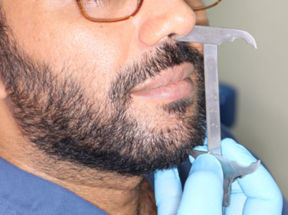
With the development of guided bone regeneration technology, researchers have mixed two or more materials to play a complementary role. To date, most studies have focused on the mixed use of autologous bone and xenogeneic bone substitute materials. The reason why this method has a better clinical effect than simple guided bone regeneration is that the inclusion of autologous bone particles brings bone-stimulating growth factors and active osteoblasts to the implanted area, which makes them rapidly vascularized. The 1:1 ratio of sausage bone grafting technology advocated by Urban has been confirmed in numerous clinical studies to have a better and more reasonable effect on repairing the bone augmentation width of the knife-edge alveolar ridge [8, 10, 14]. A comparative test between sausage bone grafts and GBR [7] revealed that the horizontal bone increment of the former was 5.3 ± 2.3 mm after 6 months of healing, whereas the horizontal bone increment of the latter was only 2.7 ± 1.8 mm. In our study, we used the sausage bone grafting technique. A total of 22 implants were included in the experimental group, and the retention rate of the implants was 100% after 1–2 years of reexamination after loading. The average bone width before surgery was 3.72 ± 0.94 mm, 11.57 ± 1.44 on the day after bone grafting, and 8.86 ± 1.37 mm after 6 months of healing. Similarly, Silvio et al. [9], reported that the average horizontal bone width increased by 5.03 ± 2.15 mm from the level of the alveolar crest up to 7 months after bone graft healing.
Regarding the resorption rate during repair of the knife-shaped alveolar ridge, the bone absorption rate during the healing of the bone graft area in this study was 0.33 ± 0.16, which was slightly higher than that reported in the study of Helene M et al. (0.29) [7]. One reason was the oppressive influence of muscle activities and the physiological structure and limitations of the materials used; another reason is that bone reconstruction is a continuous dynamic process, and therefore, slight variations may be related to small differences in the recovery time during healing.In addition, maxillary teeth were included in most of the above studies, and previous studies have shown that there is a difference in the healing speed of the upper and lower jaws [15]. Previous studies have shown good bone augmentation results; the researchers hypothesized that these bone gains are due to the composition of the granular grafts used in the study (a 1:1 mixture of granular autografts and DBBM), as xenografts slow the uptake of autografts and promote an increase in volume [14, 16]. Moreover, a meta-analysis reported that bone resorption via xenografts was lower (11.6%) than that via autografts alone [17]. In addition, Amorfini's and Gultekin's studies reported similar results, with a positive correlation between graft volume and graft absorptivity when a 1:1 mixture of granular autografts and DBBM was used [18, 19]. However, unlike the other studies, the present study did not reveal a correlation between graft volume and graft material absorption during healing [20]. We believe that the bone formation volume of the jaw is not completely proportional to the amount of bone graft, and the calculation of the resorption rate is closely related to the amount of bone graft on the day of surgery; thus, the resorption rates calculated by various experts differ. On the basis of the results from the above mentioned Urban study, the absorption rate is likely to be stable regardless of the amount of transplanted material [7].
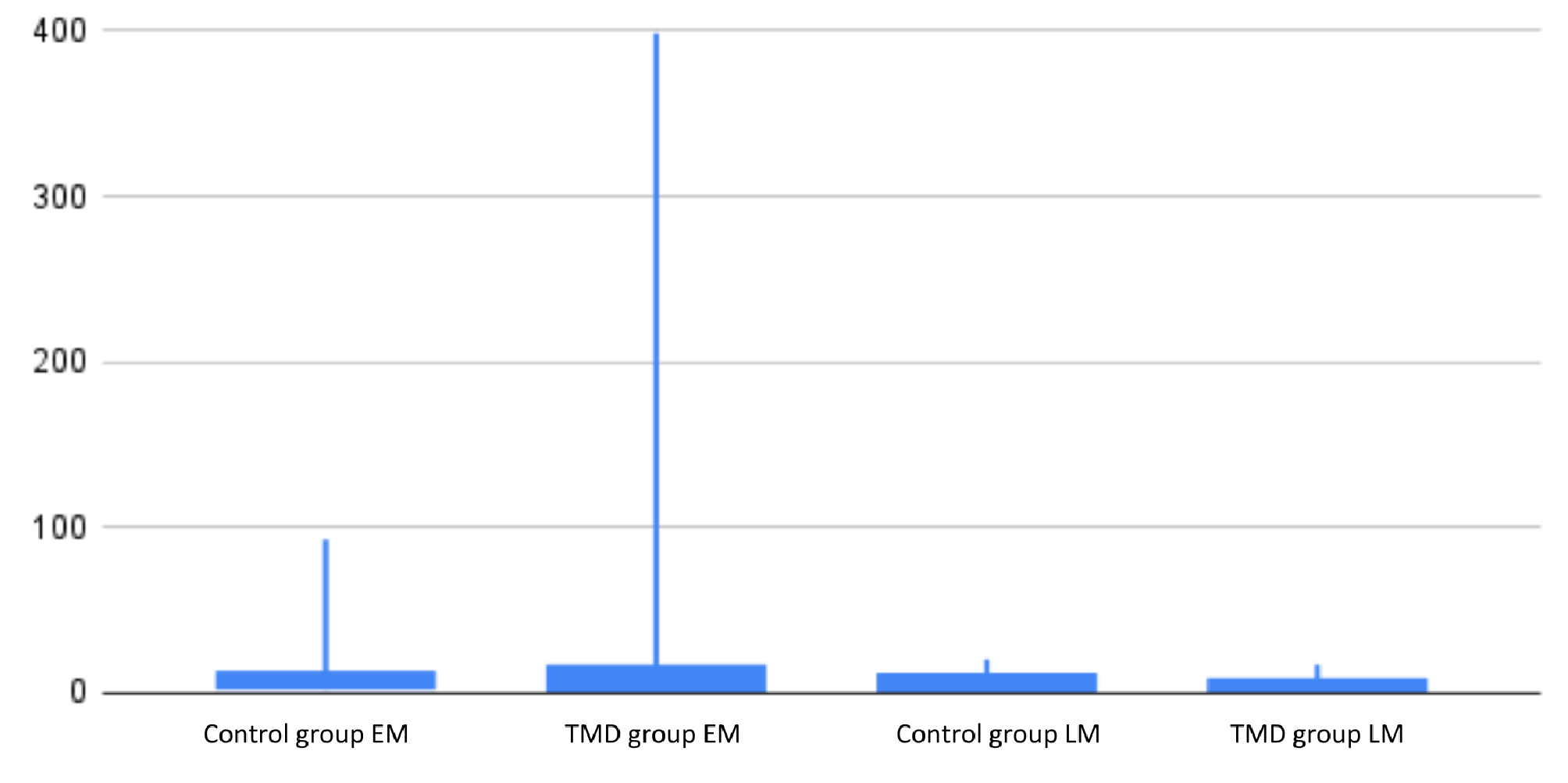
Both the dynamic stability of bone and bone remodeling are joint effects of osteogenic activity and osteoclastic activity, or they balance each other, or one side is more active. Numerous studies have confirmed that the RANK/RANKL/OPG system plays an important regulatory role in alveolar bone metabolism and remodeling and that the expression level and site affect bone resorption and bone formation [21,22,23]. RANKL plays an important role in the maturation and activation of osteoclasts.The only cell surface receptor activator of RANKL RANK. OPG, a soluble decomposing receptor, is an inhibitor of RANKL, and the two proteins have opposite effects on bone turnover. OPG binds to the activator of the RANKL receptor and reduces the binding of RANKL to RANK, thereby inhibiting the osteoclast process and promoting osteogenesis. In the study of orthodontic tooth movement, Yamaguchi, M et al. reported that RANK-RANKL plays a central role in bone resorption [24]. In addition, H.Tanaka et al. confirmed that the expression of RANKL/OPG was the key factor in bone formation and bone resorption during bone remodeling [25]. Therefore, the total RANKL/OPG ratio may be a reasonable indicator that explains the results of studies on the biological activity of these biomarkers as indicators of tissue change. Multiple studies have shown that a high RANKL/OPG ratio promotes osteoclast activity and subsequent bone loss, suggesting a tendency for bone resorption in patients; a low RANKL/OPG ratio leads to osteogenesis and subsequent bone remodeling, suggesting a tendency toward osteogenesis in patients [26,27,28]. Reducing RANKL expression or regulating the RANKL/OPG ratio is an effective strategy to prevent alveolar bone resorption and promote bone formation. In tissue engineering therapy, the RANKL/RANK/OPG signaling pathway can be directly and locally promoted to induce osteogenic differentiation to enhance the formation of new bone [29, 30]. In vitro experiments, Qu Z, et al.and Toledano-Osorio M, et al. all found that stimulating osteoblast-like cells on the surface of the implant led to alkaline phosphatase activity and osteocalcin production of cells were increased, while OPG/RANK ratio increased significantly [31, 32]. In the mouse experiment, Akiyama et al. inhibited the expression level of RANKL and increased the expression level of OPG in mouse primary osteoblasts to prevent cell differentiation into osteoclasts and bone resorption [33]. In the clinical study, the RANKL/OPG ratios for healthy implants were 0.10 ± 0.09 and 0.08 ± 0.08 for smoking and non-smoking [34]. The dates are lower than the results of our study. On the one hand, we did not compare smoking and non-smoking groups, and there is a dependent relationship between ratios and the amount and length of the smoking habit. On the other hand, the small sample size could be a limitation of our study. At present, most studies report results at the diseased sites of periodontal, and there is little information about the biomarkers in healthy peri-implant fluids. In addition, studies have shown that a certain amount of bone remodeling occurs at the beginning of the load on the alveolar crest in the neck of the implant restoration, as do changes in cytokine levels [35]. Therefore, in our study, the experimental group was compared with the control group and natural teeth. The results indicated that the RANKL level in the gingival crevicular fluid of the two groups appeared similar to that of the natural teeth, while the OPG level in the experimental group tended to be higher than that in the control group and natural teeth, although this difference was not statistically significant. The RANKL/OPG ratios of the implants in both groups all tended to be lower than that in the natural teeth,while the ratio of the implants in the experimental group was lower than that in the control group, although the differences were not statistically significant. Therefore, we believe that the osteogenesis process may still be ongoing after 1 to 2 years of loading of the implant in the bone graft area. We also compared implants loaded for 12–18 months with those loaded for 18–24 months and found that the OPG levels were slightly higher around implants that were loaded for a shorter period and were greater than those of healthy natural teeth. The RANKL concentration and the RANKL/OPG ratio were slightly lower. In addition, we also quantitatively measured the levels of several inflammatory factors around the implants and performed routine clinical periodontal examinations [36]. There was no significant difference between the experimental group and the control group. The stability of the surrounding soft and hard tissue was very similar to that of healthy natural teeth when the control group implant was loaded for 12–24 months. However, the levels of IL- 1β, IL- 6, TNF-ɑ and MMP- 8 in the experimental group were slightly greater than those in the control group, which indicated that the peri-implant bone remodeling in the experimental group was to some extent more active than that at the control group. Therefore, we hypothesized that osteogenic activity was more active in 18 months after loading, and decreased after 18 months, but it was still weakly exist. Further investigation should include expanding the sample size, increasing the measurement time point for the gingival crevicular fluid, and increasing genetic testing to confirm the diagnosis and prognosis.


Comments (0)