
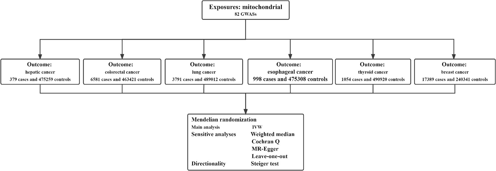
Remember me


As shown in Fig. 2A, in the MR study investigating the causal relationship between mitochondria and hepatic cancer, we observed a negative correlation with hepatic cancer for “39S ribosomal protein L34, mitochondrial” (OR = 0.796, 95% CI = 0.642–0.987, P = 0.037), “Mitochondrial fission regulator 1” (OR = 0.824, 95% CI = 0.685–0.991, P = 0.039), and “Mitochondrial import inner membrane translocase subunit TIM14” (OR = 0.778, 95% CI = 0.621–0.974, P = 0.028), indicating them as protective factors. Conversely, “[Pyruvate dehydrogenase (acetyl-transferring)] kinase isozyme 2, mitochondrial” (OR = 1.545, 95% CI = 1.040–2.294, P = 0.031), “Steroidogenic acute regulatory protein, mitochondrial” (OR = 1.173, 95% CI = 1.033–1.331, P = 0.014), and “Coiled-coil-helix-coiled-coil-helix domain-containing protein 10, mitochondrial” (OR = 1.171, 95% CI = 1.045–1.311, P = 0.007) exhibited a positive correlation with hepatic cancer, indicating them as risk factors. The Steiger test confirms that “Coiled-coil-helix-coiled-coil-helix domain-containing protein 10, mitochondrial” (Steiger P value < 0.001) is an upstream factor for hepatic cancer. Apart from these 6 exposure factors with a causal relationship with hepatic cancer, no significant associations were found with the other 76 mitochondrial-related exposures.
Fig. 2
MR Forest plot and leave-one-out analyses of the causal relationship between mitochondria and hepatic cancer. A. The results from Mendelian randomization analysis using the IVW method, MR-Egger regression and Weighted median method. B. Coiled-coil-helix-coiled-coil-helix domain-containing protein 10, mitochondrial. C. Mitochondrial import inner membrane translocase subunit TIM14. D. 39 S ribosomal protein L34, mitochondrial. E. Mitochondrial fission regulator 1. F. [Pyruvate dehydrogenase (acetyl-transferring)] kinase isozyme 2, mitochondrial. G. Steroidogenic acute regulatory protein, mitochondrial. OR, odds ratio. CI, confidence interval
Causal effects of mitochondria on colorectal cancerAs illustrated in Fig. 3A, in the MR study investigating the causal relationship between mitochondria and colorectal cancer, we observed a negative correlation with colorectal cancer for “Phenylalanine-tRNA ligase, mitochondrial” (OR = 0.946, 95% CI = 0.901–0.994, P = 0.028), “Malonyl-CoA decarboxylase, mitochondrial” (OR = 0.877, 95% CI = 0.785–0.979, P = 0.019), and “Mitochondrial import inner membrane translocase subunit TIM14” (OR = 0.896, 95% CI = 0.821–0.977, P = 0.013), indicating them as protective factors. Conversely, “Methylmalonyl-CoA epimerase, mitochondrial” (OR = 1.107, 95% CI = 1.021-1.200, P = 0.014) and “Coiled-coil-helix-coiled-coil-helix domain-containing protein 10, mitochondrial” (OR = 1.072, 95% CI = 1.028–1.118, P = 0.001) exhibited a positive correlation with colorectal cancer, indicating them as risk factors. Through Cochran Q test, heterogeneity was detected in the causal relationship between “Malonyl-CoA decarboxylase, mitochondrial” (P value = 0.041) and colorectal cancer. The Steiger test confirms that “Phenylalanine-tRNA ligase, mitochondrial” (Steiger P value = 0.029) and “Coiled-coil-helix-coiled-coil-helix domain-containing protein 10, mitochondrial” (Steiger P value < 0.001) are upstream factors for colorectal cancer. Apart from these identified 5 exposure factors with a causal relationship with colorectal cancer, no significant associations were found with the other 77 mitochondrial-related exposures.
Fig. 3
MR Forest plot and leave-one-out analyses of the causal relationship between mitochondria and colorectal cancer. A. The results from Mendelian randomization analysis using the IVW method, MR-Egger regression and Weighted median method. B. Coiled-coil-helix-coiled-coil-helix domain-containing protein 10, mitochondrial. C. Mitochondrial import inner membrane translocase subunit TIM14. D. Phenylalanine-tRNA ligase, mitochondrial. E. Methylmalonyl-CoA epimerase, mitochondrial. F. Malonyl-CoA decarboxylase, mitochondrial. OR, odds ratio. CI, confidence interval
Causal effects of mitochondria on lung cancerAs shown in Fig. 4A, in the MR study investigating the causal relationship between mitochondria and lung cancer, we observed a negative correlation with lung cancer for “Succinate dehydrogenase assembly factor 2, mitochondrial” (OR = 0.942, 95% CI = 0.894–0.992, P = 0.024), indicating it as a protective factor. Conversely, “Superoxide dismutase [Mn], mitochondrial levels” (OR = 1.054, 95% CI = 1.011–1.099, P = 0.013) exhibited a positive correlation with lung cancer, indicating it as a risk factor. The Steiger test confirms that “Superoxide dismutase [Mn], mitochondrial levels” (Steiger P value = 0.014) is an upstream factor for lung cancer. Apart from these 2 identified exposure factors with a causal relationship with lung cancer, no significant associations were found with the other 80 mitochondrial-related exposures.
Fig. 4
MR Forest plot and leave-one-out analyses of the causal relationship between mitochondria and lung cancer. A. The results from Mendelian randomization analysis using the IVW method, MR-Egger regression and Weighted median method. B. Superoxide dismutase [Mn], mitochondrial levels. C. Succinate dehydrogenase assembly factor 2, mitochondrial. OR, odds ratio. CI, confidence interval
Causal effects of mitochondria on esophageal cancerAs depicted in Fig. 5A, in our MR study, we observed a positive correlation between “Lon protease homolog, mitochondrial” (OR = 1.152, 95% CI = 1.006–1.320, P = 0.039) and esophageal cancer, indicating it as a risk factor. The Steiger test confirms that “Lon protease homolog, mitochondrial” (Steiger P value = 0.007) is an upstream factor for esophageal cancer. Besides this exposure factor with a causal relationship with esophageal cancer, no significant associations were found with the other 81 mitochondrial-related exposures.
Fig. 5
MR Forest plot and leave-one-out analyses of the causal relationship between mitochondria and esophageal cancer. A. The results from Mendelian randomization analysis using the IVW method, MR-Egger regression and Weighted median method. B. Lon protease homolog, mitochondrial. OR, odds ratio. CI, confidence interval
Causal effects of mitochondria on thyroid cancerAs depicted in Fig. 6A, in the MR analysis of mitochondria and thyroid cancer, we found a negative correlation with thyroid cancer for “Iron-sulfur cluster assembly enzyme ISCU, mitochondrial” (OR = 0.770, 95% CI = 0.625–0.949, P = 0.014) and “Coiled-coil-helix-coiled-coil-helix domain-containing protein 10, mitochondrial” (OR = 0.885, 95% CI = 0.785–0.997, P = 0.045), indicating them as protective factors. Conversely, “Diablo homolog, mitochondrial” (OR = 1.218, 95% CI = 1.034–1.434, P = 0.018) and “Persulfide dioxygenase ETHE1, mitochondrial” (OR = 1.200, 95% CI = 1.001–1.438, P = 0.049) exhibited a positive correlation with thyroid cancer, signifying them as risk factors. The Steiger test confirms that “Coiled-coil-helix-coiled-coil-helix domain-containing protein 10, mitochondrial” (Steiger P value < 0.001) is an upstream factor for thyroid cancer. Apart from these identified 4 exposure factors with a causal relationship with thyroid cancer, no significant associations were found with the other 78 mitochondrial-related exposures.
Fig. 6
MR Forest plot and leave-one-out analyses of the causal relationship between mitochondria and thyroid cancer. A. The results from Mendelian randomization analysis using the IVW method, MR-Egger regression and Weighted median method. B. Coiled-coil-helix-coiled-coil-helix domain-containing protein 10, mitochondrial. C. Diablo homolog, mitochondrial. D. Persulfide dioxygenase ETHE1, mitochondrial. E. Iron-sulfur cluster assembly enzyme ISCU, mitochondrial. OR, odds ratio. CI, confidence interval
Causal effects of mitochondria on breast cancerAs shown in Fig. 7A, in the MR study investigating the causal relationship between mitochondria and breast cancer, we identified “ADP-ribose pyrophosphatase, mitochondrial” (OR = 0.963, 95% CI = 0.930–0.997, P = 0.033), and “Cytochrome c oxidase subunit 8A, mitochondrial” (OR = 0.959, 95% CI = 0.928–0.990, P = 0.011) as protective factors, showing a negative correlation with breast cancer. Conversely, “39S ribosomal protein L34, mitochondrial” (OR = 1.069, 95% CI = 1.010–1.132, P = 0.021), “Pyruvate carboxylase, mitochondrial” (OR = 1.071, 95% CI = 1.019–1.127, P = 0.007), “rRNA methyltransferase 3, mitochondrial” (OR = 1.031, 95% CI = 1.006–1.057, P = 0.015), and “Cytochrome c oxidase assembly factor 3 homolog, mitochondrial” (OR = 1.067, 95% CI = 1.016–1.121, P = 0.009) were identified as risk factors, showing a positive correlation with breast cancer. The Steiger test confirms that “ADP-ribose pyrophosphatase, mitochondrial” (Steiger P value < 0.001), “rRNA methyltransferase 3, mitochondrial” (Steiger P value < 0.001), and “Cytochrome c oxidase subunit 8A, mitochondrial” (Steiger P value < 0.001) serve as upstream factors for breast cancer. Besides these identified 6 exposure factors with a causal relationship with breast cancer, no significant associations were found with the other 76 mitochondrial-related exposures.
Fig. 7
MR Forest plot and leave-one-out analyses of the causal relationship between mitochondria and breast cancer. A. The results from Mendelian randomization analysis using the IVW method, MR-Egger regression and Weighted median method. B. Cytochrome c oxidase assembly factor 3 homolog, mitochondrial. C. Cytochrome c oxidase subunit 8 A, mitochondrial. D. 39 S ribosomal protein L34, mitochondrial. E. ADP-ribose pyrophosphatase, mitochondrial. F. Pyruvate carboxylase, mitochondrial. G. rRNA methyltransferase 3, mitochondrial. OR, odds ratio. CI, confidence interval
Sensitivity analysisAs shown in Table 2, there is heterogeneity in the causal relationship between “Malonyl-CoA decarboxylase, mitochondrial” and colorectal cancer. In the remaining IVW calculations, a P value < 0.05 was obtained, with Cochran Q test P value > 0.05, MR-Egger intercept test P value > 0.05, and no significant impact from individual SNPs. Therefore, the other causal relationships are statistically significant, demonstrating no heterogeneity or horizontal pleiotropy, and the assessment results are reliable. Detailed sensitivity analysis and directionality test results are provided in Table 2. The leave-one-out analyses results illustrating the causal relationships between mitochondria and six types of cancer through MR are presented in Figs. 2, 3, 4, 5, 6 and 7.
Table 2 The results of the Cochran Q test, Egger intercept test, and Steiger test were used to assess the causal relationship between mitochondria and six types of cancer
Comments (0)