
Remember me


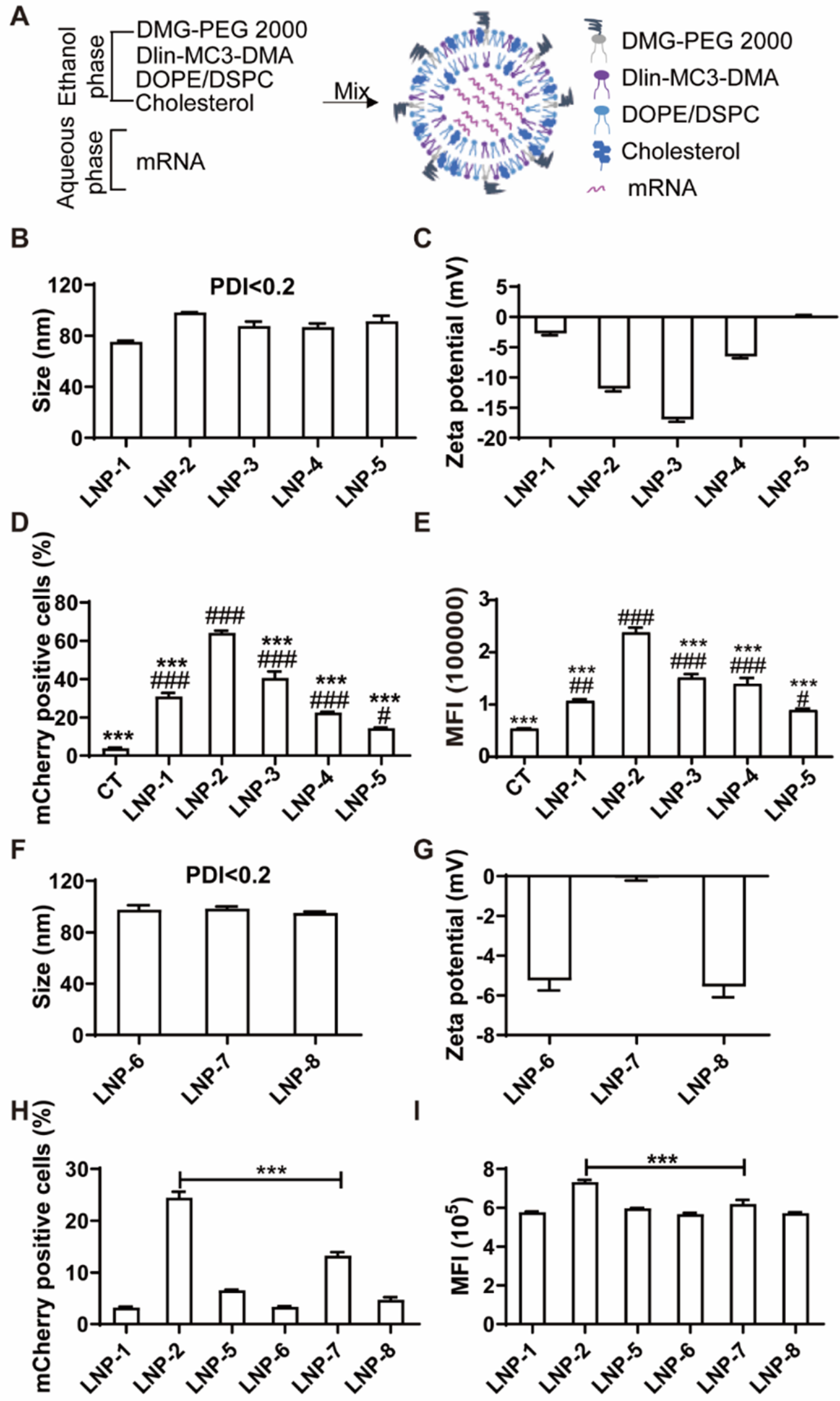

Current FDA-approved LNPs for mRNA vaccines and siRNA delivery utilize DSPC as a helper lipid. However, DSPC was originally designed for siRNA delivery and exhibits low dissociation efficiency for long-chain nucleotides like mRNA [22]. Structurally, DOPE contains a cis-double bond in each aliphatic tail, enhancing mRNA release, whereas DSPC has fully saturated tails that strongly associate with RNA. Mechanistically, DSPC adopts a cylindrical phase, forming stable lipid bilayers, while DOPE adopts a conical shape that transitions to a hexagonal conformation, promoting membrane fusion [23]. To address this limitation, we developed DOPE-containing LNPs, which are more conducive to mRNA release. These LNPs consist of four lipid components: DLin-MC3-DMA, cholesterol, DOPE, and DMG-PEG 2000 (Fig. 1A).
Fig. 1
Characterization of LNPs with different phospholipids at different mole ratios and their delivery efficiency of mCherry mRNA in ECs. (A) Schematic of assembly of LNP. Sizes (B) and Zeta potentials (C) of DOPE containing LNPs at different mole ratios with mCherry mRNA. Quantification of mCherry positive cells (D) and Mean fluorescence intensity (E) of HUVECs transfected with DOPE-LNP encapsulated with mCherry mRNA at 24 h post-transfection. # P < 0.05, ## p < 0.01, ## P < 0.001 versus control; *** P < 0.001 versus LNP-2 group. Sizes (F) and zeta potentials (G) of LNPs with DSPC at different mole ratios. Quantification of mCherry positive cells (H) and mean fluorescence intensity (I) of HUVECs transfected with DOPE or DSPC containing LNPs encapsulated with mCherry mRNA at 6 h post-transfection. *** P < 0.001 versus LNP-2 group. Data are presented as the mean ± SEM
To optimize the lipid composition for mRNA delivery, we synthesized a series of LNPs encapsulated with mCherry mRNA (LNP-1 through LNP-5) with different mole ratios of the four lipids (Fig. S1). Each formulation had a polydispersity index (PDI) of less than 0.2, indicating a monodisperse distribution. Hydrodynamic diameters ranged from 80 to 100 nm (Fig. 1B), and zeta potentials ranged from − 17 to 0 mV (Fig. 1C).
To assess transfection efficiency, HUVECs were treated with LNP1-5 loaded with mCherry mRNA. All formulations significantly enhanced mCherry expression compared to free mRNA delivery (Fig. 1D and E). Notably, LNP-2 achieved the highest transfection efficiency, with 65% of cells being mCherry-positive (Fig. 1D) and a mean fluorescence intensity (MFI) of 240,000 compared to 40,000 in the control group (Fig. 1E). Representative FACS histograms are shown in Fig. S2A. These findings indicate that LNP-2 provides an optimal mole ratio for efficient mRNA delivery.
Given the structural characteristics of DOPE, which supports effective mRNA release, we hypothesized that DOPE-containing LNPs would outperform traditional DSPC-containing LNPs in delivering mRNA. To test this, we synthesized DSPC-containing LNPs (LNP-6 through LNP-8) with mole ratios corresponding to LNP-1, LNP-2, and LNP-5, respectively (Fig. S1). All DSPC-LNPs encapsulated with mCherry mRNA were monodisperse with PDI values < 0.2, had diameters between 95 and 99 nm (Fig. 1F), and exhibited zeta potentials from − 6 to 0 mV (Fig. 1G). Additionally, encapsulation efficiency measured by RiboGreen assays showed that 5 out of the 8 LNP formulations achieved over 80% encapsulation efficiency (Fig. S1).
When comparing transfection efficiency in HUVECs, DOPE-containing LNPs (LNP-1, LNP-2, and LNP-5) consistently showed superior delivery performance relative to their DSPC counterparts (LNP-6, LNP-7, and LNP-8) (Fig. 1H and I). Specifically, LNP-2 showed the highest percentage of mCherry-positive cells (25%) and MFI (73,000), markedly outperforming LNP-7, its DSPC equivalent. Representative FACS histograms are shown in Fig. S2B. These data support that DOPE-containing LNPs are more efficient for mRNA delivery than DSPC-containing LNPs.
Based on these findings, LNP-2 (35:46.5:16:2.5 ratio of DLin-MC3-DMA, cholesterol, DOPE, and DMG-PEG 2000) was selected for further experiments. mRNA-encapsulated LNP-2 formulations were named LNP@mCherry, LNP@Fluc, and LNP@SDF-1α based on the specific mRNA payload (mCherry, firefly luciferase, or SDF-1α, respectively).
DOPE-LNPs are low cytotoxic, stable, and highly efficient for mRNA delivery in vitroTransmission electron microscopy (TEM) images confirmed the spherical morphology of both empty LNP-2 (Fig. 2A) and mCherry mRNA-loaded LNP-2 (Fig. 2B), with diameters consistently around 100 nm. The particles exhibited good dispersion in suspension without notable aggregation.
Fig. 2
The shape, cytotoxicity, stability of LNPs and their delivery efficiency of mCherry mRNA in ECs at different times. TEM images of empty LNP-2 (A) and mcherry mRNA encapsulated LNP-2 (B). Scale bar: 50 nm. (C) Cytotoxicity was examined by CCK-8 assay of HUVECs incubated with empty LNP-2 at various concentrations for 48–72 h. (D) Sizes of LNPs encapsulated with mRNA kept at 4 °C up to 60 days post assembly. Quantification of mCherry positive cells (E) and mean fluorescence intensity (F) of HUVECs transfected with LNP@mCherry or naked mCherry mRNA for 6, 18, or 36 h. ** P < 0.01, *** P < 0.001 versus Free mCherry group. Data are presented as the mean ± SEM
To evaluate the cytotoxicity of LNP-2, HUVECs were treated with empty LNP-2 at concentrations from 12.5 to 100 µg/mL for both 48 and 72 h. A CCK-8 assay indicated that cell viability was unaffected compared to the control group, suggesting that DOPE-LNPs possess low cytotoxicity (Fig. 2C).
The stability of LNP@mRNA was assessed by monitoring particle size at 4 °C over 15, 30, and 60 days post-assembly. No significant change in particle size (~ 100 nm) was observed, confirming the high stability of these formulations over time (Fig. 2D).
Naked mRNA has been consistently shown to have a limited ability to cross the cell membrane due to its negative charge [24]. To evaluate the mRNA delivery efficiency of LNPs in vitro, we assessed the mCherry fluorescent signal at 6, 18, and 36 h post-transfection of mCherry mRNA. Free mCherry mRNA was used as a control, and LNPs alone served as the vehicle control. Consistent with previous reports, free mCherry mRNA generated a weak fluorescent signal, detectable only at 36 h post-transfection, in contrast to the robust signal observed in the LNP group. At each time points, LNP-2 demonstrated a substantially increased percentage of mCherry-positive cells and higher mean fluorescence intensity (MFI) compared to free mRNA (Fig. 2E and F). Representative FACS histograms are displayed in Fig. S3.
Overall, these results confirm that LNP-2 is non-cytotoxic, highly stable, and can deliver mRNA with high efficiency in vitro.
LNP@SDF-1α promotes endothelial network formation, proliferation, migration and CXCR4 positive cell adhesionThe hydrodynamic diameter of LNP@SDF-1α was measured at 110 nm (Fig. 3A), with a zeta potential of -10 mV (Fig. 3B). To evaluate the delivery efficiency of SDF-1α mRNA, HUVECs were transfected with LNP@SDF-1α for 24 h, resulting in significantly increased mRNA levels (Fig. 3C), protein expression (Fig. 3D) and secretion into culture supernatant (Fig. 3E) as assessed by RT-PCR, western blot, and ELISA assay compared to the control.
Fig. 3
The angiogenesis, migration, and adhesion effects of LNP@SDF-1α in vitro. Sizes (A) and zeta potentials (B) of LNP@SDF-1α. SDF-1α mRNA was delivered to HUVECs for 24 h. (C) SDF-1 mRNA levels. (D) Protein levels. (E) The secreted SDF-1α protein levels in the culture supernatant examined by ELISA. (F) Proliferation of HUVECs. (G) Quantification of numbers of junctions and vessel length using AngioTool software. (H) Representative images of tube formation of HUVECs treated with LNP or LNP@SDF-1α. (I) Representative images of cell migration after scratch. (J) Quantification of cell migration in I. (K) The adhesion of THP-1 cells to HUVECs. (L) Quantification of cell adhesion in K. (M) The adhesion of THP-1 cells to HUVECs. HUVECs were treated with 0.4 µg LNP-delivered SDF-1α for 6 h. THP-1 cells were pre-incubated with 10 µM CXCR4 antagonist AMD3100 8HCl for 30 min, then added to ECs. (N) Quantification of cell adhesion in M.* P < 0.05, ** P < 0.01, *** P < 0.001. Data are presented as the mean ± SEM
To compare the expression efficiency of mRNA and plasmid, the SDF-1α level in the supernatant was tested by ELISA. The SDF-1α protein level expressed by mRNA was significantly higher than that of plasmid (Fig. S4).
The effect of LNP@SDF-1α on the survival of HUVECs was examined by CCK-8 kit. As shown in Fig. 3F, LNP@SDF-1α could slightly promote the proliferation of HUVECs. To assess angiogenic potential, HUVECs were transfected with LNP@SDF-1α or control LNP for 6 h before being subjected to a Matrigel tube formation assay. As shown in Fig. 3G and H, LNP@SDF-1α-treated cells displayed a marked increase in tube formation compared to controls. Quantitative analysis using AngioTool software confirmed significant increases in the number of junctions and total vessel length (Fig. 3G) in the LNP@SDF-1α group.
The migration capacity of HUVECs post-transfection with LNP@SDF-1α was evaluated by scratch assay. LNP@SDF-1α treatment significantly enhanced migration, with wound closure increasing from 42.3 to 60.9% (Fig. 3I and J).
Additionally, the effect of LNP@SDF-1α on the adhesion of human CXCR4-positive THP-1 cells to ECs was investigated. LNP@SDF-1α-treated HUVECs demonstrated enhanced monocytic cell adhesion (Fig. 3K), which was further increased in a dose-dependent manner with escalating concentrations of SDF-1α mRNA (Fig. 3L). To further determine whether the SDF-1α-induced enhancement is dependent on its receptor CXCR4, we applied CXCR4 antagonist AMD3100 8HCl and observed a significant reduction in cell adhesion (Fig. 3M and N). This results confirm that SDF-1α-mediated promotion of cell adhesion is CXCR4-dependent.
LNP@SDF-1α enhances angiogenesis in the matrigel plug assayTo assess the angiogenic potential of LNP@SDF-1α in vivo, we utilized a mouse Matrigel plug assay (Fig. 4A).
Fig. 4
The angiogenesis effects of LNP@SDF-1α in vivo. (A) Schematic of the Matrigel plugs assays. (B) Bright-field image of LNP@SDF-1α induced angiogenesis in Matrigel plugs. (C) Representative images of hematoxylin and eosin staining of Matrigel plugs with the indicated treatment. (D) Quantification of blood vessels containing red blood cells. (E) Immunofluorescent staining of Matrigel plugs. (F) Quantification of the number of vessels in E. * P < 0.05, *** P < 0.001. Data are presented as the mean ± SEM
HUVECs treated with LNP@SDF-1α were embedded in Matrigel, while LNP-treated cells served as controls. These Matrigel plugs were then injected subcutaneously into 8-week-old C57BL/6 mice, and harvested five days post-injection for analysis. Matrigel plugs containing HUVECs transfected with SDF-1α mRNA displayed a darker red color, indicative of increased blood content and vascular density compared to control plugs (Fig. 4B).
HE staining revealed a significant increase in the number and size of blood vessels containing red blood cells in LNP@SDF-1α-treated HUVEC plugs compared to the control group (Fig. 4C and D). Immunofluorescent staining for CD31 further confirmed enhanced vascularization in the LNP@SDF-1α-treated group (Fig. 4E and F).
These results conclusively demonstrate that LNP@SDF-1α promotes angiogenesis in vivo, supporting its potential as an effective therapeutic for vascular regeneration.
Intramuscular injection of LNP@SDF-1α enhanced the angiogenesis, blood perfusion, and preserved limb function in murine hindlimb ischemia modelTo evaluate the local expression dynamics of LNP-mediated mRNA delivery in vivo, we administered an intramuscular injection of LNP encapsulating 4 µg of Fluc mRNA into mice and monitored whole-body bioluminescence at 4, 24, and 48 h post-injection. While 4 µg of naked Fluc mRNA was also examined to determine their delivery efficiency. Bioluminescence served as an indicator of Fluc expression. At 4 h post-injection (Fig. 5A and B), we only observed robust bioluminescence at the local injection site treated with LNP encapsulated Fluc mRNA, indicating that LNP successfully delivered mRNA, which was rapidly translated into functional protein in vivo. Bioluminescence was also detectable at 24 and 48 h post-injection, suggesting a sustained expression for at least two days. Free Fluc mRNA without LNP exhibited minimal bioluminescence, similar to LNP group.
To directly compare the delivery efficiency of recombinant protein, plasmid, and mRNA forms of SDF-1α, we injected SDF-1α protein, naked SDF-1α plasmid, and SDF-1α mRNA encapsulated by LNP into the gastrocnemius. After 4 h, the plasma was collected, and SDF-1α protein levels were measured by ELISA. In both the protein and plasmid injection groups, SDF-1α levels remained comparable to those in untreated control plasma (Fig. 5C). This aligns with previous reports indicating that recombinant proteins are rapidly degraded in tissues, while plasmid-driven expression in vivo is delayed and relatively low. In contrast, SDF-1α mRNA administration resulted in robust protein expression, highlighting its potential for rapid and efficient therapeutic delivery.
Fig. 5
Therapeutic effects of LNP@SDF-1α in the lower limb ischemia model at 21 d. (A) The expression of Free Fluc mRNA or Fluc mRNA delivered by LNP in vivo. (B) Quantification of fluorescence intensity in A. (C) The SDF-1α protein level in the plasma from mice treated with SDF-1α protein, plasmid or mRNA. * versus the control group; # versus the plasmid group. (D) Schematic of the treatment of the lower limb ischemia. (E) Laser Speckle Contrast Imaging of the ischemic hindlimb on Days 0, 3, 7, 14, and 21 after injury. (F) Quantification of the blood flow perfusion ratio of the ischemic limb compared with the nonischemic limb (n = 5). (G) Limb lost score in ischemic limb. (H) Scoring for the physiological status of ischemic limbs at Day 21. * P < 0.05, ** P < 0.01, *** P < 0.001. # P < 0.05. Data are presented as the mean ± SEM
We next assessed the therapeutic efficacy of LNP@SDF-1α on revascularization in a mouse hindlimb ischemia model (Fig. 5D). LNP@SDF-1α or control LNP was injected intramuscularly immediately after surgery and again three days later. Laser Speckle Contrast Imaging was used to monitor tissue reperfusion (Fig. 5E), showing significantly faster blood flow recovery in ischemic limbs treated with LNP@SDF-1α compared to controls, particularly at 7, 14, and 21 days post-surgery (Fig. 5F). The limb loss score and quantitative scores of limb salvage (Fig. 5G and H) were also markedly improved in the LNP@SDF-1α group. A higher Limb loss score indicates more adverse symptoms. These findings suggest that injection of LNP@SDF-1α can improve blood perfusion and help preserve limb function in the ischemic limbs of mice.
To examine angiogenesis, immunofluorescence staining was performed on gastrocnemius muscles at 21 days post-surgery. Capillary (vessels contain CD31+ EC layer) density and arteriole (vessels contain CD31+ EC layer and α-SMA+ smooth muscle layer) density were significantly elevated in the LNP@SDF-1α treated group compared to controls (Fig. 6A). Quantitative analysis further confirmed that LNP@SDF-1α significantly increased the number of capillaries (Fig. 6B) and arterioles (Fig. 6C).
Fig. 6
LNP@SDF1α significantly promote angiogenesis at Day 21 following hindlimb ischemia. (A) Representative images of immunofluorescent staining with α-SMA (red), CD31 (green), and DAPI for nuclei (blue) on Day 21 after surgery. Quantification of the capillary (B) and artery density (C) in A. (D) The relative mRNA levels of human SDF-1α, mouse Vegf-a, and Hif-1α in muscles tested by RT-PCR. (E) The mouse angiogenesis array detected multiple analytes in muscles. (F) Quantification of selected angiogenesis-related proteins by gray analysis. * P < 0.05, ** P < 0.01, *** P < 0.001. Data are presented as the mean ± SEM
Additionally, LNP@SDF-1α treatment upregulated Vegf and Hif-1α mRNA levels, and notably, human SDF-1α mRNA was still detectable at 21 days post-injection (Fig. 6D).
We also examined angiogenesis-related protein expression using an angiogenesis array (Fig. 6E and F). LNP@SDF-1α treatment elevated pro-angiogenic factors such as angiopoietin-1 (Ang-1), hepatocyte growth factor (HGF), placental growth factor-2 (PIGF-2), and VEGF, along with an increase in matrix metalloproteinase-3 (MMP-3) levels. In contrast, the anti-angiogenic factor serpin F1 was significantly downregulated.
Histological examination of major organs (liver, spleen, and kidney) revealed no significant toxicity associated with LNP or LNP loaded with mRNA, supporting the favorable safety profile of our newly developed DOPE-LNP and modified mRNA (Fig. S5).
Comments (0)