Accurate estimation of PMI is challenging, especially when dealing with cadavers in advanced stages of decomposition [6]. The decomposition process affects the body’s external appearance, making it difficult to estimate even an approximate PMI. Additionally, factors such as temperature, humidity, and the presence of insects and scavengers can accelerate or slow down the decomposition process, further complicating the estimation of PMI [31, 32]. According to this knowledge, we designed an in corpore study under controlled ambient conditions with the body protected from insects and animals involved in decomposition. The main goal of this study was to determine the dynamics of the decrease in the fraction of viable chondrocytes from the knee of a deceased person. We assumed that regularity in this dynamic might place cartilage as a useful tool to establish a new method for the estimation of PMI. Previous pilot studies showed that knee cartilage is the most reliable source of cartilage because it is not affected by rapid decomposition, especially between temperatures around 11 °C and room temperature [15]. Other cartilaginous tissues such as the nasal septum, thyroid cartilage, larynx, trachea, spine, ribs, bronchi, shoulders and hips are expected to be more rapidly affected by decomposition due to their close contact to the commensal bacteria reservoir [16].
Analyzing the cartilage form 35 donors, with 131 samples, we confirmed earlier conclusions about chondrocytes’ ability to live a long period after the death of a host [11,12,13,14,15,16, 33]. We have observed that chondrocytes from knee cartilage can be found alive after more than two months (Figs. 3 and 4).
Furthermore, our study on whole bodies in the late PMI showed that the fraction of living chondrocytes decreased over time using all methods (CVA, FCN, FCC), which supports the conclusions from previous studies [11,12,13,14,15,16, 33]. Although fraction of viable chondrocyte’s decreased postmortem, we observed that confident interval around these estimated non-linear association was wide, even in a controlled environmental condition.
Given the relatively small number of measurements, there was considerable uncertainty in the estimation of viability curves, particularly in the initial and final weeks of our study, as can be seen from the light blue areas presented in the figures (Figs. 3 and 4).
Therefore, an extensive study with a larger number of donors and identification of potential factors that are associated with the individual’s variability, such as chronic and prior to death diseases, medications, body mass index, lifestyle etc., might narrow the confident interval in order to create a usable method for the estimation of PMI based on a chondrocyte’s viability.
Based on results from this study, we designed predictive curves for each assay, with 95 % prediction interval, to show how this method could be used in everyday practice (Fig. 4); for an individual with e.g. FCC = 0.75, the predicted PMI is 6 to 17 days, for an individual with FCC = 0.5, the predicted PMI is 13 to 26 days, while for an individual with FCC = 0.25, the predicted PMI is 21 to 30 days.
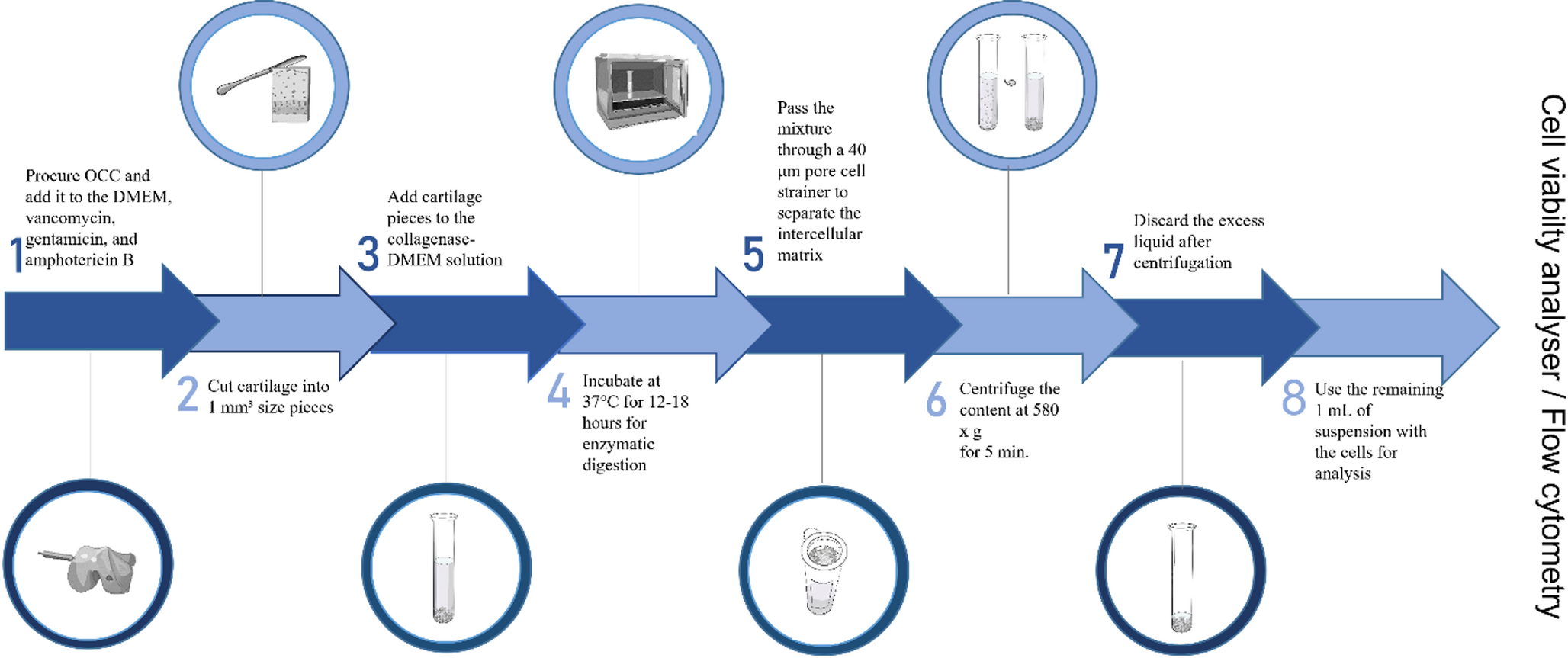
To determine the fraction of live chondrocytes we used two instruments; CVA and FC. CVA provides automatic and cost/time-effective viable-cell counting, using a trypan blue stain. Since the dye penetrates through damaged membranes of cells, the cells that become stained blue are regarded as non-viable, whereas non-stained cells are assumed to be intact and are considered viable [34].
Flow cytometry (FC) is a modern and advanced diagnostic technique that plays a crucial role in the diagnosis, monitoring of therapies, and assessment of various diseases. It is a gold standard for rapid multi-parametric analysis of single-cells in solutions [20, 35]. When analyzing cells from solid tissues, mechanical and enzymatic degradation is needed and the preparation of single-cell suspension is a crucial part of FC to avoid cell death and aggregation. This procedure should yield a cell population with high viability, minimal cell debris or aggregates, and preserved cell surfaces for FC to be effective [16, 25]. Cell viability can be determined by morphological changes or by changes in membrane permeability. According to this, DNA-binding dyes such as 7-AAD are used as dead cell discriminators in daily routines at laboratories worldwide [36]. Disadvantages of this staining can be gathered debris from dead cells in the sample, shaped to look like live cells and in that way misinterpreted. In this study, in order to avoid false results, we used RedDot™1 to identify all chondrocytes with nuclei (FCN) and 7-AAD, to distinguish live/dead chondrocytes (FCC) among them.
According to the results obtained from this study, FCC is the most promising method for predicting the PMI, followed by CVA and FCN. However, the opportunity of analyzing a huge number of samples in a short time with minimal user involvement requirement and low-cost performance [16], support the CVA to be used in further studies/routine work.
Comparing the results of different chondrocyte assays, we noticed that CVA showed a sharp decline of the curve during the first 30 days, while later it plateaued at about 30 % (Fig. 3). The inconsistency of this observation compared to the results obtained by FC and previous studies is likely due to potential CVA technical issues. One of them might be during the preparation of single-cell suspension such as cell debris and cell aggregation and the other might be due to the set device parameters. Even though in this study CVA was set to specific parameters for chondrocytes (Table 1), software capabilities of the instrument still require better adjustment of input parameters to differentiate cells within clusters allowing a more extensive major control for analyzing samples for each cell line [34].
It is well known that aging impairs the ability of cells to maintain metabolic homeostasis, leading to disruption of cartilage and reducing regenerative capacity. These age-related alterations are recognized as major contributors to the development of osteoarthritis and the progressive deterioration of articular cartilage [37]. In this study, we observed no age-related differences in the ratio of live chondrocytes when assessed using the FC method. An exception was noted in the CVA method, where an even higher proportion of live chondrocytes was found in older donors (Table 3). We hypothesize that this unexpected result may be due to the inclusion of only macroscopically intact cartilage into this study, which could account for the preservation of chondrocyte viability in older individuals. It is also important to note that our assessment focused on the ratio of live/dead chondrocytes, rather than the absolute number of viable cells. Although the total number of chondrocytes in healthy cartilage decrease with age, the rate of postmortem chondrocyte degradation may not necessarily be age dependent. High viability detected by CVA in older donors may be due to technical issues rather than to biological differences.
The use of human osteochondral allografts (OCA) has become a vital option for orthopedic surgeons to treat chondral and osteochondral lesions primarily of the knee, as well as lesions in the ankle, hip, and shoulder. Fresh OCA tissues are ideally harvested from healthy donors, with healthy gross articular cartilage, aged 14 to 50 years within 24 h of death [38]– [39]. Although cartilage from older individuals is generally more susceptible to degenerative changes, our findings suggest that, when macroscopically intact, cartilage from elderly donors may still possess adequate chondrocyte viability and could thus be considered as a potential source for OCA transplantation.
Most of the previous studies for the determination of the PMI did not show differences between males and females when calculating PMI [40,41,42,43,44,45,46]. In addition, none of the three cell viability assays (CVA, FCN, FCC) used in this study showed significant differences in the fraction of live chondrocytes between males and females which suggests that rapidity of postmortem body changes is not associated with sex.
The ability of chondrocytes to remain viable for extended periods after the death of individuals has attracted the attention of forensic experts in the last few years. In this study, we were able to generate prediction curves with a 95 % confidence interval based on postmortem chondrocyte viability, but the confidence interval was wide in some periods and as such is not currently applicable in daily practice. Larger studies could gather more information about individual differences. Furthermore, it is essential to conduct studies in different post-mortem environmental conditions such as temperature, burial, and precipitation to evaluate the effects of external factors on cellular degradation [47]. Incorporating specific correction factors, like those used in the Henssge nomogram, could contribute to the development of more precise, quantitative methodologies for estimating late PMI based on chondrocytes viability.


Comments (0)