Our research clearly indicated that DECT has the potential to differentiate between specific shotgun pellet materials. This preliminary study suggests that DECT could be a valuable tool in the radiological examination of patients with shotgun-related gunshot injuries. Our findings align with previous research on differentiating projectile materials using DEI values and the extended CT scale (Gascho et al. 2020; Winklhofer et al. 2014). We thus believe that this method could be beneficial for not just clinical forensic cases but also when reconstructing the course of events for postmortem cases when several shotgun pellet types have been used.
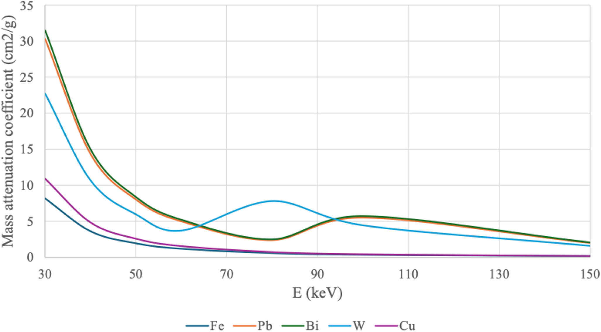
Steel and copper shots exhibited distinct DEI values. In contrast, lead, tungsten, and bismuth had similar DEI values to each other and the separation between these materials was not feasible. It can thus be concluded that although DECT can categorize pellet materials, it clearly exhibits some limitations with exact material detection. The high Wilcoxon test p-value of 0.869 for Bi and Pb and the comparable box plots illustrated that these two pellet materials exhibited particularly similar DEI values. This observation aligns well with the nearly identical mass attenuation coefficients of the materials (Fig. 1). Furthermore, differentiating tungsten from bismuth and lead was also observed to be difficult, consistent with the similarities in their mass attenuation coefficients (Fig. 1). Moreover, the strong overall attenuation of these high-atomic-number materials (ZBi = 83, ZPb = 82, ZW = 74) compared to those materials with lower atomic numbers (ZFe = 26, ZCu = 29) may also contribute to the similarities in DEI values. To improve tungsten differentiation, the DECT protocol could be optimized so that the low-kV acquisition would produce an effective energy of approximately 80 keV, where tungsten exhibits deviating mass attenuation properties compared to Pb and Bi (Fig. 1). However, given the strong attenuation of these materials, care must be taken to prevent signal saturation.
Based on our results, DECT can be potentially utilized for ferromagnetic projectile material detection as already demonstrated by Winklhofer et al. (2014). In this study, steel pellets had distinct DEI values compared to other pellet materials; only copper pellets had relatively similar values to steel ones. This finding is very useful as steel and lead are the most common pellet materials and copper is among the least common ones. Distinguishing between lead, tungsten, and bismuth that are all not ferromagnetic is not a major disadvantage as especially the reliable detection of a ferromagnetic steel shot would be particularly useful in living shotgun injury victims with an urgent need for MRI. More detailed studies are, however, required to create the basis for a reliable methodology to be further utilized in clinical use.
Several studies have recognized the need to increase our understanding of shotgun-related gunshot injuries (Mushtaque et al. 2012). Further studies describing the radiological examination of shotgun wounds in various regions of the body are warranted (Cañas et al. 2007). This would be particularly important for a further understanding of the tendency of pellets to migrate between tissues creating internal damage. In addition, it would be important to be able to distinguish potentially toxic pellet materials from less harmful ones.
We believe that our preliminary study provides an important step for the radiological analysis of shotgun pellets and could be beneficial for both clinical and postmortem cases. We could demonstrate the potential of DECT in foreign material identification and especially categorization and confirm the findings of previous studies that are mainly concentrating on bullet and jacket materials (Gascho et al. 2020; Winklhofer et al. 2014). Our promising results encourage the future use of DECT when victims of shotgun-related incidents are examined in clinical and forensic contexts.
One of the main strengths of the presented approach is its high level of automation for material detection. Our approach was automated and computationally efficient, and could thus be performed on standard computing hardware, such as a laptop. The simplicity and speed of this workflow would make it an accessible tool for broader use in clinical and research settings. However, further studies are required to confirm and develop the methodology presented in this preliminary study. It should also be noticed that the radiation dose of DECT should be optimized for the material identification task in a diagnostic context, in which the as low as reasonably achievable (ALARA) principle should be followed.
The utilized protocol and radiation dose, in particular, warrants a separate discussion. The selected radiation dose aligns with the 2018 American College of Radiology CT Accreditation Program adult diagnostic reference level of 25 mGy (ACR-AAPM-SPR 2024), which was the most recent document at the time of measurements. For improved traceability to this standard and other similar material quantification studies, such as (Jacobsen et al. 2019), we selected this radiation dose level. However, the most recent diagnostic reference levels in Finland are substantially lower than this value. As an example, the dose reference level for abdominal CT in Finland is 12 mGy (CTDIvol).
Our main worry for the present study was the possibility that high noise may hide some of the smaller shrapnels due to the association between image noise and low contrast detectability. However, based on the results, the attenuation from the pellets is so strong that substantially lower doses could be utilized for reliable detection. Furthermore, for diagnostically relevant materials, e.g., iodine, there is strong evidence that the radiation dose level does not have a significant effect on the material quantification accuracy of dual-energy CT excluding diagnostically very low doses (Sartoretti et al. 2022; Jiang et al. 2021). Thus we expect that this dose-independence holds also for the pellet materials. This research question could be examined as a follow-up study.
As a potential limitation, the pellet materials were not confirmed by chemical analysis, but the information was obtained from the ammunition manufacturers. It is possible and even probable that some of the materials were not purely made of just one substance but alloys of two or more substances. This would be the case especially with tungsten pellets as they would be prone to shatter in the pure form. In addition, the pellets of steel shot are not actually steel but made of soft iron. This doesn’t, however, play a significant role from the radiological point of view.
One potential limitation in the pellet analysis could be the sensitivity to the partial volume and beam hardening effects, particularly in relation to the size of the projectile being analysed. Preliminary tests were conducted to explore the impact of pellet size on DEI results. For instance, only minor differences in DEI values were observed between 3 mm and 4 mm steel pellets. As the size of the projectile decreases further, partial volume effect may lead to inaccurate DEI values. This is especially relevant when dealing with very small fragments, where the blending of tissue and material boundaries can skew the results (Guo et al. 2011). However, in shotgun wounds, this should not produce major problems as smallest common pellets are 2 mm in diameter, and in general, pellets tend to remain intact.
On the other hand, the presence of larger metallic objects, such as more sizable pellets or shrapnel, may introduce beam hardening artifacts. However, shotgun pellets rarely are more that 4.5 mm in diameter and in extreme cases their diameter may be up to 8 mm. Beam hardening, known to cause inaccuracies in HU measurements, could similarly affect DEI values. Consequently, beam hardening may introduce another variable that could complicate the interpretation of DEI data (Kanatani et al. 2021). Future research should systematically investigate the influence of both small and large fragment sizes on DEI accuracy to better understand the impact of partial volume and beam hardening before considering the diagnostic adoption of the presented method.
Another important consideration relates to the thresholding method used for pellet segmentation. A limitation of our automated segmentation approach is the use of a fixed 2000 HU threshold, which may not be sufficient to distinguish shotgun pellets from other dense foreign objects in clinical or forensic imaging, such as dental prostheses or metallic implants. While this threshold was effective in our controlled gelatine block environment, real-life cases may require additional criteria. Our segmentation code is flexible in this regard. It identifies each connected component individually, which would allow users to inspect and verify each segmented object separately. The workflow also allows integration of additional shape or size-based filtering, which can help restrict the analysis to fragments with dimensions and morphology consistent with shotgun pellets, reducing the inclusion of irrelevant objects in diagnostic and forensic imaging.
The medico-legal significance of this study can be highlighted from several perspectives. First, in medico-legal systems where autopsies cannot necessarily be performed on an immediate basis, postmortem CT may constitute a means of providing the police with relevant pieces of information rapidly, for example, on the material of shotgun pellets associated with a homicide case. Second, in mass disasters, the resources for performing full medico-legal autopsies in a timely manner may be limited. While providing an efficient method to document findings, CT scans may also be used for the rapid extraction of data regarding, for example, projectile materials deposited in a victim. Third, in routine medico-legal autopsies, manually searching for pellets in tissues may be time-consuming and not all pellets can be retrieved despite all efforts. This may increase the possibility of error, in particular in cases where several pellet types have been used together, and not all can be analyzed. Fourth, among living patients with shotgun injuries, imaging often is the only means of evaluating the properties of the pellets embedded in the body.


Comments (0)