Ethics statement
This study was approved by the Ethics Committee of Huashan Hospital, Fudan University (KY2024-1020). All the experiments involving animals were approved by the Animal Care and Use Committee of Fudan University (No. 202410048S).
Human NPC isolation and culture
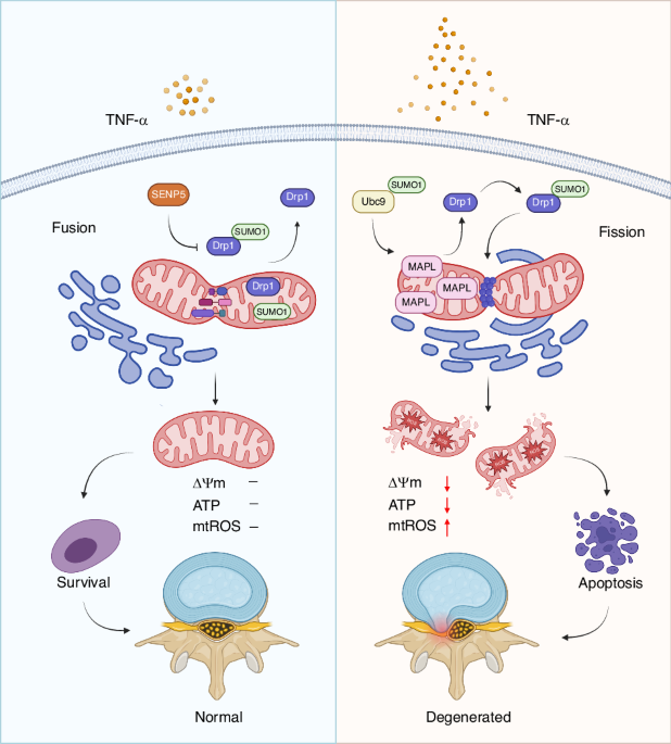
Primary NPCs were isolated from IVD tissues obtained from 26 patients diagnosed with Hirayama disease who underwent anterior cervical discectomy and fusion. Magnetic resonance imaging (MRI) was used to evaluate the degree of IVDD according to the Pfirrmann grading system. To guarantee that only healthy, non-degenerated IVD tissues were included, the Pfirrmann grade I classification was strictly adhered to during sample selection. The NP tissues were cut into pieces and digested with 0.2% collagenase II (Thermo Fisher Scientific, USA) for 8 h at 37 °C. The released cells were collected by centrifugation for 5 min at 500 × g. The NPCs were then resuspended and cultured in Dulbecco’s modified Eagle’s medium (DMEM) supplemented with 15% foetal bovine serum (FBS) and maintained in an incubator at 37 °C with 5% CO2. The culture medium was changed every three days. NPCs at the second passage were used for further experiments. To mimic NP degeneration in vitro the NPCs were treated with 10 ng/mL TNF-α for 24 h.
RNA sequencing
Total RNA was extracted from the control and TNF-α groups using TRIzol reagent (Invitrogen, USA). After being qualitatively checked, the mRNA was enriched with oligo (dT)-coated magnetic beads and fragmented for reverse transcription into cDNA with random primers. Then, the cDNA fragments were purified, terminally modified and ligated to sequencing adapters. The ligation products were size selected by gel electrophoresis, amplified by PCR and used for cDNA library preparation. Libraries were sequenced using the HiSeq X Ten system (Illumina, USA). The raw sequencing data were processed and mapped to the human reference genome using STAR software. Genes that were differentially expressed (a P value cut-off of 0.05 and a fold-change cut-off of 2) between the two groups were analysed using the DESeq2 package.
RNA interference and plasmid transfection
The plasmids pcDNA3.1-MAPL, pcDNA3.1-SENP5, WT-Drp1, 4KR-N Drp1 (K532R, K535R, K558R, K568R), 4KR-C Drp1 (K594R, K597R, K606R, K608R) and 8KR Drp1 (K532R, K535R, K558R, K568R, K594R, K597R, K606R, K608R) were constructed by GeneChem (Shanghai, China). A set of siRNAs was designed and synthesised by GeneChem (Shanghai, China). The sequences used were as follows: Homo si-MAPL: 5′-AGGAGCTGTGCGGTCTGTTAAAGAA-3′; si-NC (MAPL): 5′-GATACTTGACTAGGAAACCCACACA-3′; Homo si-Drp1: 5′- GCTGTTTCTAAAGTTTCCCAGTATA-3′; si-NC (Drp1): 5′- CCATGGTCTAATTGTCACATCATGT-3′; Homo si-SENP5: 5′- GCATCAGGTTGTAGTTGCATCTTTA-3′; and si-NC (SENP5): 5′- CAGAGAACTTACAGTGTACAACATA-3′. The NPCs were transfected with plasmids or siRNAs using Lipofectamine 3000 (Invitrogen, USA) according to the manufacturer’s instructions.
Western blot analysis
NPCs were lysed in RIPA buffer (AspenTech, China) on ice, and the protein concentration was quantified using a BCA protein assay kit (AspenTech, China). The isolated proteins were separated by sodium dodecyl sulfate‒polyacrylamide gel electrophoresis (SDS‒PAGE) (Millipore, USA). The proteins were subsequently transferred onto polyvinylidene fluoride (PVDF) membranes (Millipore, USA), which were subsequently blocked with 5% milk. Primary antibodies were added, and the membranes were incubated overnight at 4 °C. After incubation with horseradish peroxidase-conjugated secondary antibodies, the membranes were visualised with chemiluminescence reagents (AspenTech, China) and an imaging system. The primary antibodies used were as follows: anti-MAPL (16133-1-AP, 1:1 000, Proteintech), anti-SENP5 (ab58420, 1:500, Abcam), anti-SUMO1 (67559-1-Ig, 1:1 000, Proteintech), anti-cleaved caspase-3 (AF7022, 1:500, Affinity Biosciences), anti-Bax (#2772, 1:1 000, Cell Signalling Technology), anti-Bcl-2 (1:1 000, ab196495, Abcam), anti-Drp1 (#8570, 1:1 000 (IB) or 1:100 (IP), Cell Signalling Technology), anti-VDAC1 (ab15895, 1:3 000, Abcam) and anti-β-actin (TDY051, 1:10 000, TDY Biotech).
Coimmunoprecipitation
After the indicated treatments, the NPCs were washed twice with precooled PBS and lysed with NP-40 lysis buffer (AspenTech, China) supplemented with a 1% protease inhibitor cocktail (AspenTech, China) and 1% PMSF (AspenTech, China) on ice for 30 min. After centrifugation at 12 000 r/min for 15 min at 4 °C, the supernatant was collected, and the protein concentration was determined with a BCA protein assay kit (AspenTech, China). The same amount of protein from each group was added to 10 μL of anti-Drp1 antibody (Cell Signalling Technology, USA) and incubated with Protein A/G magnetic beads (Bio-Rad, USA) at 4 °C overnight. The immunoprecipitate was eluted with SDS‒PAGE loading buffer at 95 °C for 10 min and analysed by Western blot analysis.
Immunofluorescence staining
NPCs were fixed with 4% paraformaldehyde for 20 min, permeabilised with 0.5% Triton X-100 for 15 min and blocked with 5% bovine serum albumin for 30 min. The samples were then incubated with the primary anti-Drp1 antibody (1:100) at 4 °C overnight. After three washes with PBS, the samples were incubated with the corresponding secondary antibodies for 40 min in the dark. The nuclei were stained with DAPI (C1006; Beyotime, China) for 20 min. Finally, representative images were acquired using a fluorescence microscope (Olympus, Japan) and fluorescence confocal microscope (Zeiss, Germany).
Apoptosis assay
NPC apoptosis was evaluated by flow cytometry and Annexin V-FITC/PI staining. An apoptosis analysis kit (BD Biosciences, USA) was used to stain the NPCs. Briefly, the NPCs were incubated with 5 μL of Annexin V-FITC and 5 μL of PI for 15 min in the dark after being resuspended. The rate of apoptosis was analysed using a flow cytometer (Beckman Coulter, USA).
Mitochondrial membrane potential (ΔΨm) assessment
The ΔΨm was measured using a JC-1 Assay Kit (Beyotime, China). NPCs were incubated with JC-1 staining working solution for 20 min at 37 °C. The samples were subsequently washed twice with JC-1 buffer solution. After being resuspended, the stained NPCs were analysed via flow cytometry. The ratio of JC-1 aggregates to monomers was estimated as an indicator of alterations in the mitochondrial membrane potential.
Measurement of mitochondrial reactive oxygen species (mtROS) and cellular ROS levels
Cellular ROS and mtROS levels were examined with a ROS assay kit (Beyotime, China) and MitoSOX Red (Thermo Fisher Scientific, USA), respectively. NPCs were stained with MitoSOX Red for 30 min or DCFH-DA for 20 min at 37 °C in the dark. The cells were then washed three times with PBS. The NPCs were observed under a fluorescence microscope (Olympus, Japan) to detect mtROS. Flow cytometry (Beckman Coulter, USA) was used to measure the cellular ROS level.
Mitotracker staining
MitoTracker Green (Beyotime, China) and MitoTracker Red CMXRos (Beyotime, China) were used to stain mitochondria according to the manufacturer’s instructions. Briefly, after the media were discarded, the NPCs were incubated with MitoTracker Green working solution or MitoTracker Red working solution at 37 °C for 30 min. A fluorescence confocal microscope (Zeiss, Germany) was used to capture representative images.
Transmission electron microscopy
NPCs were fixed with 2.5% glutaraldehyde for 1 h at 4 °C. The cells were then fixed with 1% osmium tetroxide for 2 h at 37 °C. The samples were subsequently dehydrated and embedded in Epon 812 (Shell Chemicals, USA). Ultrathin sections were cut at thicknesses of 60–70 nm with an ultramicrotome (Leica, Germany) and stained with uranyl acetate and lead citrate. Images were obtained with a transmission electron microscope (Hitachi, Japan).
Animal model and intradiscal injection
We used adeno-associated virus (AAV)-mediated overexpression and knockdown of MAPL in animal experiments. The AAVs were purchased from GeneChem (Shanghai, China). Twenty-four Sprague‒Dawley rats (male, 8 weeks old) were randomly assigned to four groups (n = 6): the AAV-NC group, IVDD + AAV-NC group, IVDD + AAV-MAPL group, and IVDD + AAV-shMAPL group. The rats were anaesthetised via an intraperitoneal injection of 2% pentobarbital (40 mg/kg). The experimental level IVD at Co7/8 was located by digital palpation of the coccygeal vertebrae. A 20-gauge needle was used for vertical puncture at the centre of the disc through the AF and into the NP. The depth of penetration was approximately 5 mm. The needle was rotated 360° and kept in the disc for 30 s. A total of 2 µL of solution containing the AAV vector, AAV-MAPL or AAV-sh-MAPL (1 × 10⁹ pfu/mL) was intradiscally injected into the centre of the NP region with a microlitre syringe once a week for four weeks to investigate the biological functions of MAPL. Finally, the rats were returned to their cages with unrestricted activity and sacrificed one month after the surgery.
MRI
Radiographic evaluation of the rat coccygeal vertebrae was performed with MRI (BioSpec 70/30 USR, Bruker, Germany) to assess signal intensity alterations and structural abnormalities in sagittal T2-weighted sequences. The severity of IVDD was classified according to the Pfirrmann grading system, a well-established clinical classification method.31
Histological staining and immunohistochemistry
The samples were collected, fixed, decalcified, dehydrated, embedded in paraffin, and cut into sections. H&E, Safranin O-fast green and Alcian blue staining were performed to analyse the degree of IVDD. Immunohistochemistry was performed to analyse the expression levels of MAPL (16133-1-AP, 1:200, Proteintech), SUMO1 (67559-1-Ig, 1:500, Proteintech), and cleaved caspase-3 (AF7022, 1:200, Affinity Biosciences). The integrated optical density (IOD) of the immunohistochemical images was quantified using ImageJ software.
Statistical analysis
The data in this study are presented as the means ± standard deviations (SDs). Three biological replicates were performed for the in vitro experiments, and six biological replicates were performed for the in vivo experiments. The statistical analyses were performed using SPSS 21.0 and GraphPad Prism 9.0 software. The data were analysed via Student’s t test for comparisons between two groups and one-way analysis of variance followed by Tukey’s test for comparisons among multiple groups. A value of P < 0.05 indicated statistical significance (*P < 0.05, **P < 0.01 or ***P < 0.001).

Comments (0)