
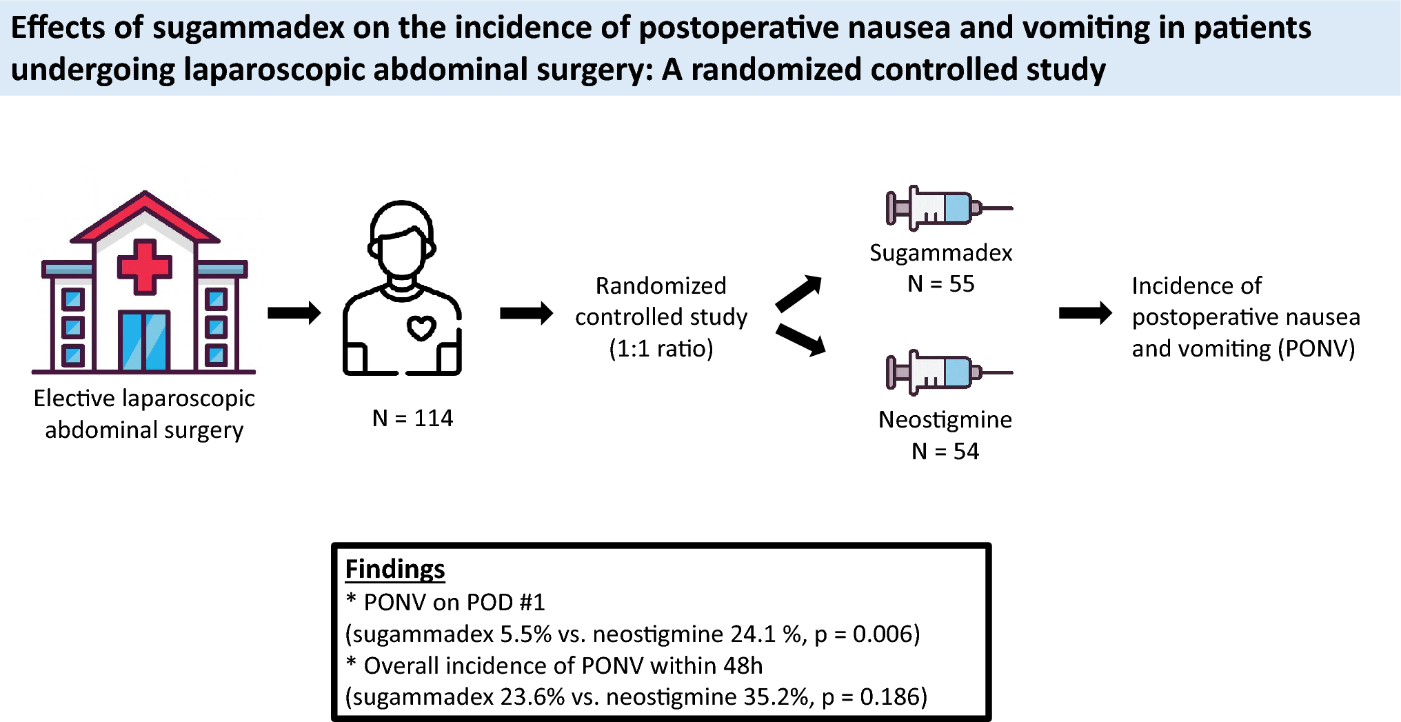
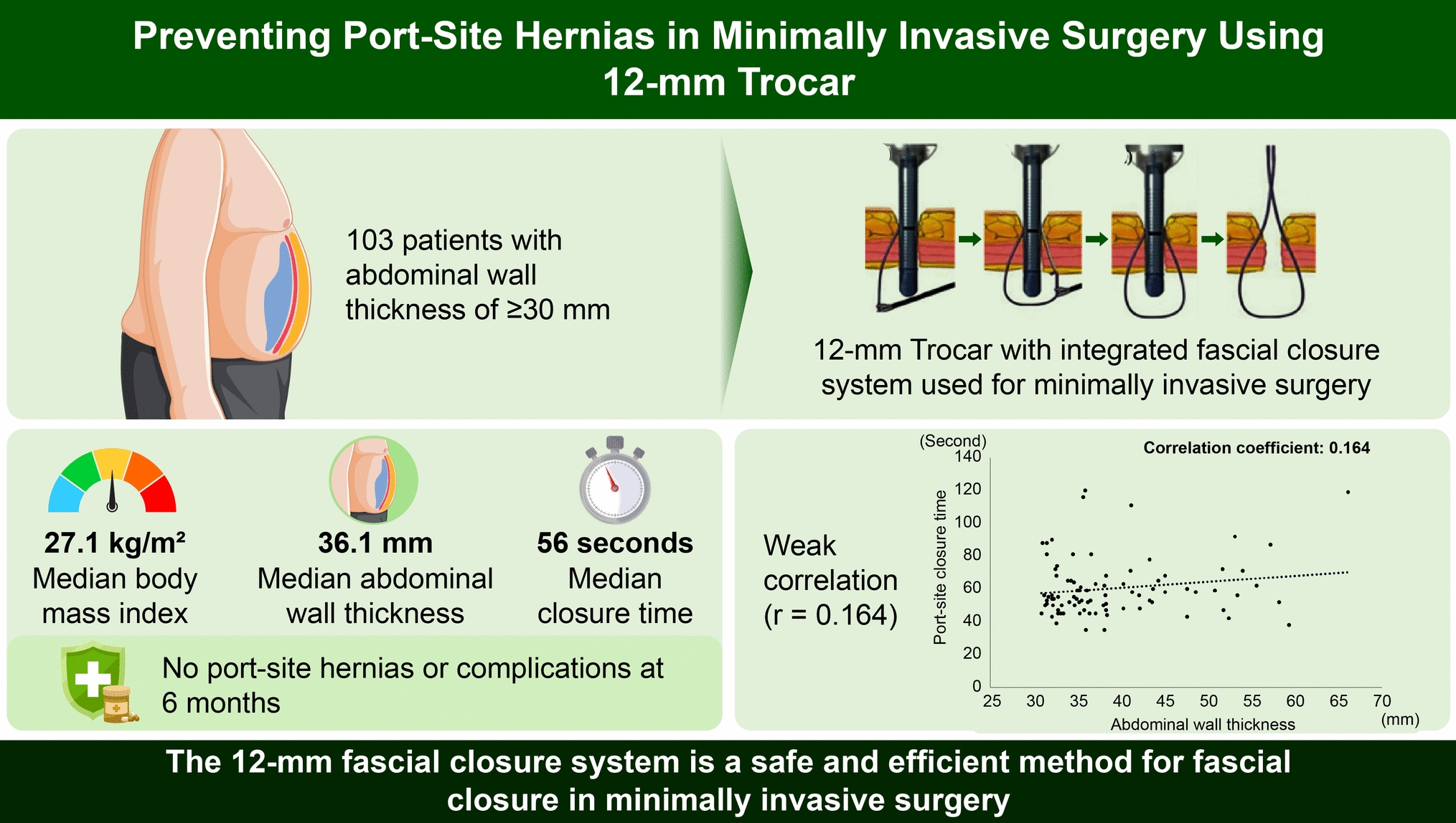
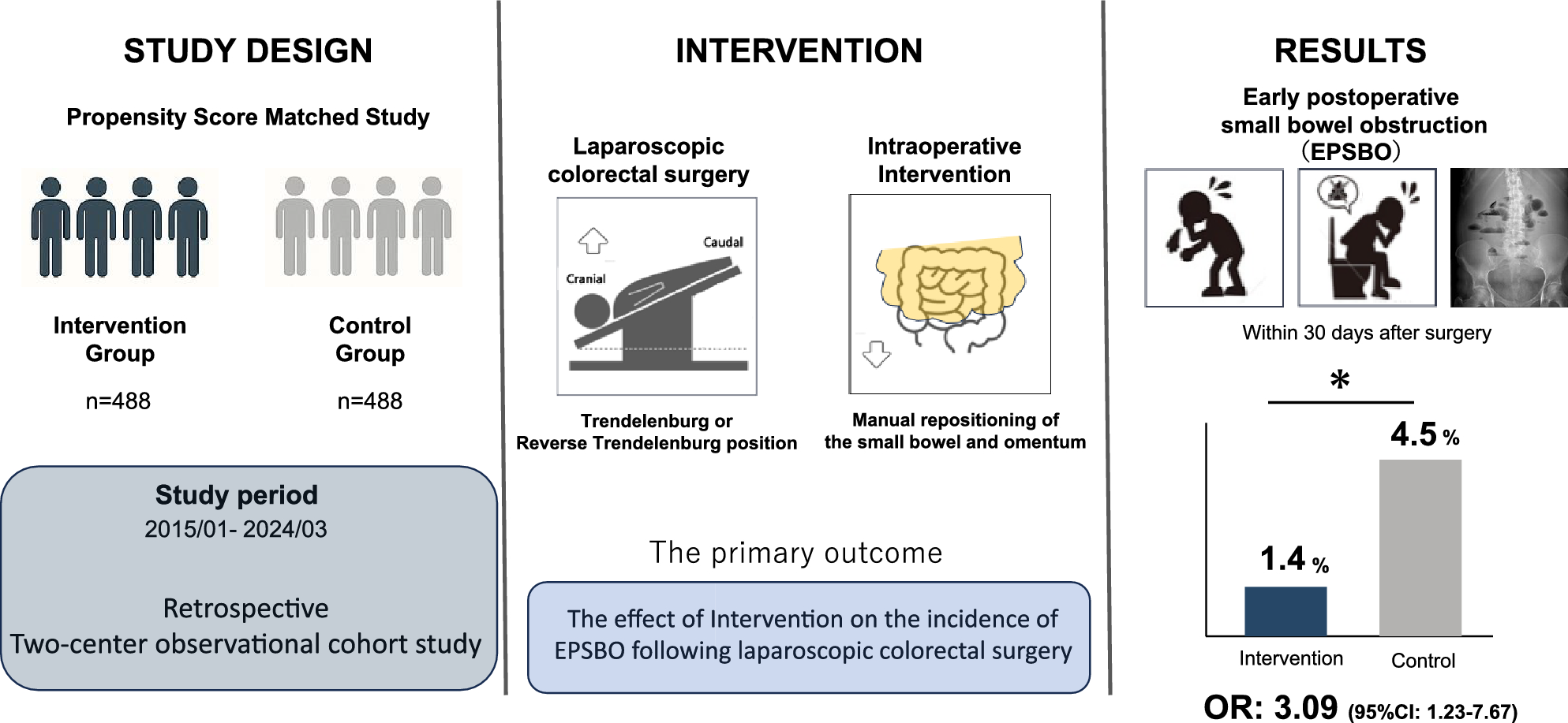
Remember me


We enrolled 90 patients, including 62 males and 28 females aged 42–83 (average 63.06 ± 8.87) years. Thirty-six of the patients successfully received single-tunnel ESTD, and 54 received double-tunnel ESTD. Each patient had one lesion in their esophagus involving at least 1/2 of the esophageal circumference. A circumferential extent of (≥ 1/2, < 3/4) was identified in 12 lesions, (≥ 3/4, < 1) in 25 lesions, and (= 1) in 53 lesions. Among the 90 lesions, 9 were found to be located in the upper esophagus, 49 were located in the middle esophagus, and 32 were located in the lower esophagus. The macroscopic type was consistent with the Paris classification type 0–IIa, 0–IIb, or 0–IIc, or a combination of these [12, 13]. Among the 90 esophageal lesions studied, 27 (30.0%) were type 0–IIa, 49 (54.4%) were 0–IIb, 3 (3.3%) were 0–IIc, and the other 11 (12.2%) were mixed type. As shown in Table 1, there was a significant difference in the degree of circumferential lesions between the two groups (P < 0.01). For whole-circumferential lesions, double-tunnel ESTD treatment was mainly adopted, whereas for noncircumferential lesions, single-tunnel ESTD treatment was the preferred approach. In terms of other clinical and endoscopic characteristics (such as age, sex, lesion location, and macroscopic classification), there was no statistically significant difference between the two groups (P > 0.05).
Table 1 Clinical characteristics and endoscopic featuresShort-term outcomesFinal pathological diagnosisA final pathological analysis revealed that 19 samples (21.1%) were finally diagnosed as high-grade intraepithelial neoplasia (HGIN), and 71 samples (78.9%) were finally diagnosed as squamous cell carcinoma (SCC). The T stage distribution among the patients is as follows: Tis (17.8%), T1a (53.3%), and T1b (28.9%). Horizontal margin involvement and vertical margin involvement were identified in 20 lesions (22.2%) and 8 lesions (8.9%), respectively. Lymphovascular invasion was identified in 4 lesions (4.4%). As shown in Table 2, there was no statistically significant difference between the single- and double-tunnel groups in terms of pathology, such as type, stage and local infiltration (P > 0.05).
Table 2 Final pathological diagnosisProcedure-related parametersEach patient had one lesion in the esophagus. All patients had completed ESTD surgery. The rates of en bloc resection, R0 resection and curative resection were 97.8%, 70.0% and 58.9%, respectively. There were 41 patients with lesion areas ≤ 20 cm2, and the remaining 49 patients had lesion areas > 20 cm2. The maximum long diameter of the seven samples was ≤ 4 cm, and those of the remaining samples were > 4 cm. The median operative time of the total sample was 100.50 (74.75–141.75) min, and the median resection speed was 0.23 (0.17–0.31) cm2/min. In terms of short-term postoperative complications, 8 patients (8.9%) experienced postoperative bleeding, and 16 patients (17.8%) experienced postoperative fever. There were no adverse events, such as perforation, cardiac mucosal laceration, or muscular damage. As shown in Table 3, the en bloc resection rate of the double-tunnel group was greater than that of the single-tunnel group, but the difference was not statistically significant (100% vs. 94.4%, P = 0.307). In terms of the R0 and curative resection rates, those of the single-tunnel group were greater than those of the double-tunnel group, but the difference was also not statistically significant (69.4% vs. 66.7%, P = 0.782; 63.9% vs. 51.9%, P = 0.259). There were significant differences in the area and maximum long diameter of the resected samples between the two groups. The overall lesion area of the double-tunnel group was significantly larger than that of the other groups, and the long diameter was significantly greater (P = 0.004, 0.03). However, there was no significant difference in operative time between the two groups [99.50 (70.75–139.50) min vs. 103.00 (79.75–147.75) min, P = 0.422]. After calculation and analysis, there was a statistically significant difference in the lesion resection speed between the two groups, with the double-tunnel group having a significantly faster resection speed than the single-tunnel group [0.25 (0.19–0.36) cm2/min vs. 0.19 (0.13–0.27) cm2/min, P = 0.012]. In terms of short-term postoperative complications, there was no statistically significant difference in incidence between the two groups (P > 0.05).
Table 3 Procedure-related parametersRisk factors for technical difficultiesTechnical difficulties were defined as the incidence of horizontal margin involvement, post-ESTD bleeding, muscular damage, and post-ESTD fever, which were 22.2% (20/90), 8.9% (8/90), 0.0% (0/90), and 17.8% (16/90), respectively. The number of events includes all events above. Among the patients with technical difficulties, each was found to experience one or more events (Fig. 2). Therefore, by analysing clinical data, the technical difficulty rate was calculated to be 40% (36/90) in the present study. All patients with adverse events were successfully treated with endoscopic therapy or conservative treatment.
Fig. 2
Patients who experienced technical difficulties
Understanding the risk factors for the technical difficulty of esophageal ESTD could help endoscopists accurately stratify lesions that are more difficult and prone to failure of en bloc resection or adverse events. Univariate and multivariate logistic regression analyses were conducted for the risk factors that may lead to technical difficulties. As shown in Table 4, the univariate regression analysis indicated that among the T stage variables, compared with Tis stage, T1a stage (OR 0.247, 95% CI 0.075–0.811, P = 0.021) was negatively correlated with the occurrence of technical difficulties, whereas there was no significant difference for the T1b stage. The multivariate regression analysis demonstrated that among the circumferential extent variables, compared with lesions with circumferential extents ranging from 1/2 to 3/4, whole-circumferential lesions (OR 11.926, 95% CI 1.380–103.04, P = 0.024) were an independent risk factor for technical difficulties. Compared with T1a stage, T1a stage (OR 0.131, 95% CI 0.030–0.569, P = 0.007) was a protective factor against the occurrence of technical difficulties. No significant differences were observed for the other risk factors.
Table 4 Univariate and multivariate analyses of risk factors for technical difficultiesLong-term outcomesEsophageal stenosisAfter the resection of extensive esophageal lesions, the surgeons adopted different preventive measures for stenosis based on the postoperative wound conditions: 13 patients (14.4%) received no special treatment for the wound, 13 patients (14.4%) received hormone injection into the wound, 14 patients (15.6%) underwent endoscopic balloon dilation, 18 patients (20.0%) had esophageal stents placed, 26 patients (28.9%) underwent autologous skin grafting, and 6 patients (6.7%) received balloon dilation combined with hormone injection (Table 5). Close follow-up was conducted after the operation. By the end of the study period, a total of 54 patients had developed stenosis, with a stenosis rate of 60%. The median time to esophageal stenosis was 59.5 (39.75–100.25) days. In the single-tunnel group, the main preventive measures for esophageal stenosis were no treatment (30.6%) and implantation of esophageal stents (25%), whereas in the double-tunnel group, the main preventive measures were autologous skin grafting (44.4%), hormone injection (16.7%), and placement of esophageal stents (16.7%). There was a significant difference in the preventive measures adopted between the two groups (P < 0.001). However, in terms of the postoperative esophageal stenosis rate, although the double-tunnel group had a higher stenosis rate (64.8% vs. 52.8%), the difference between the two groups was not statistically significant (P = 0.253). Among the 54 patients who developed esophageal stenosis, the median time to esophageal stenosis was similar in both groups (64 days vs. 56 days), and there was no significant difference (P = 0.717, Table 5).
Table 5 Comparison of postoperative management between 90 patients with single- and double-tunnel ESTDOwing to significant differences between the single-tube group and the double-tube group at the baseline level (the proportion of circumferential lesion extent and the measures for preventing stenosis), the conclusions obtained in the previous section may have been affected by selection bias. To further compare whether there were differences in the postoperative stenosis rate between the two groups, a subgroup analysis was conducted for the patients with whole-circumferential esophageal lesions (Fig. 3). As shown in Table 6, there were no significant differences between the two groups in terms of lesion area, maximum long diameter, or methods for preventing stenosis, indicating that the baseline values were comparable. The results revealed that there were no statistically significant differences between the two groups in terms of the postoperative stenosis rate or the median time of stenosis (P = 1.000, 0.649).
Fig. 3
Circumferential extent of the lesions and the treatment methods used in 90 patients
Table 6 Comparison of postoperative management between single- and double-tunnel ESTD for 53 patients with whole-circumferential lesionsSurvivalOverall survival (OS) was defined as the time from ESTD to death from any cause. Recurrence-free survival (RFS) was defined as the time from ESTD to the occurrence of distant or lymph node metastasis. Disease-specific survival (DSS) was defined as the time from ESTD to death from cancer recurrence. A total of 90 patients were included in this study, among whom 16 were lost to follow-up and 74 were successfully followed. There were 28 patients in the single-tunnel group and 46 in the double-tunnel group. Among the followed-up patients, 7 experienced successive recurrences. Five patients received corresponding treatments after recurrence (2 patients underwent ESD a second time, 2 patients underwent esophagectomy, and 1 patient received radiotherapy and chemotherapy) and are now in good condition. The other 2 patients who experienced esophageal cancer recurrence did not receive special treatment and later died with systemic metastasis. One patient died 6 months after single-tunnel ESTD, and the other died 36 months after double-tunnel ESTD. The remaining 67 patients had no recurrence (Fig. 4). As shown in Table 7, there were no statistically significant differences in the recurrence rate (P = 1.000), recurrence time (P = 0.649) or mortality rate (P = 1.000) between the two groups. K‒M survival analysis was conducted on the patients in both groups, and the results also revealed no significant differences in OS (P = 0.6242), DSS (P = 0.6242), or RFS (P = 0.6753) between the two groups (Fig. 5A–C).
Fig. 4
Recurrence status and supplementary treatment modalities of 74 follow-up patients
Table 7 Comparison of long-term outcomes between single- and double-tunnel ESTD for 74 followed-up patientsFig. 5
A‒D survival analysis of OS, DSS and RFS between the two groups
Among the 74 patients who were followed up, 45 underwent curative resection, and 29 underwent noncurative resection. To eliminate the influence of noncurative resection on tumour recurrence, Kaplan‒Meier survival analysis was further conducted on the RFS of patients who underwent curative resection. Among them, there were 20 patients in the single-tunnel group, with 2 cases of recurrence, and 25 patients in the double-tunnel group, with 2 cases of recurrence. The results revealed that there was still no statistically significant difference in RFS between the two groups (P = 0.7148, Fig. 5D).
Comments (0)