Patients
Forty-seven young patients from 41 unrelated Egyptian families with SRNS were enrolled at the Department of Pediatrics, Cairo University Children’s Hospital during the period extending from February 2021 until January 2022. SRNS was defined as the lack of complete remission after four weeks of therapy with prednisone or prednisolone at standard dose (Rovin et al. 2021). Secondary SRNS patients were excluded from the study. Patients were categorized according to the age of onset of nephrotic syndrome into congenital nephrotic syndrome (CNS) presenting within the first three months of life, infantile nephrotic syndrome (INS) presenting between 3 months and one year and childhood onset nephrotic syndrome presenting after one year.
Case notes were reviewed for personal history, age of onset and family history (parental consanguinity and/or affected family member defined as having a history of proteinuria, SRNS or renal dysfunction at a young age). Age of CKD, renal replacement therapy (RRT), if any, and clinical examination with special emphasis on extrarenal manifestations particularly neurological, ocular, auditory, skeletal and urogenital associations were all collected.
Reviewed laboratory investigations included serum albumin, cholesterol, urine analysis, urinary protein/creatinine ratio, serum creatinine and CKD stage (KDIGO 2024) as well as kidney pathology results. Sample analysis procedures, including DNA extraction, genome sequencing, bioinformatics, variant curation and protein modeling, were all performed at the Egypt Center for Research and Regenerative Medicine (ECRRM).
Genomic DNA extraction, quantification and quality assessment
DNA was extracted from K2EDTA blood tubes with the chemagic™ 360 Instrument (2024–0020,Perkin Elmer, Waltham, MA, USA) using the chemagic DNA Blood 400 Kit H96 (CMG-1091, Perkin Elmer) and the 96-chemagic rod head (PN CMG-370, Perkin Elmer). Extraction was performed using 400 µl of blood, and DNA was eluted in 100 µl following the manufacturer’s recommended protocol. Two technical replicates were generated for each biological sample. DNA was quantified by the Qubit 4 Fluorometer (Invitrogen, Carlsbad, CA, USA) using the Qubit 1 × dsDNA High Sensitivity (HS) kit (Q33230, Invitrogen). All samples were checked for impurities using the NanoDrop™ One (Thermo Scientific, Waltham, MA, USA). DNA quality was checked on the LabChip GX Touch (CLS138162, Perkin Elmer) using the Genomic DNA Assay kit (CLS760685, Perkin Elmer) with the DNA Extended Range Chip (CLS138948, Perkin Elmer) following the manufacturer’s recommendations.
Library preparation and sequencing
Library preparation for genome sequencing was performed using IlluminaTruSeq DNA PCR-free library preparation kit (20,015,962, Illumina) following the manufacturer’s instructions. Samples were normalized to 1 μg, and DNA was sheared by sonication with a focused ultrasonicator M220 (PN 500295, Covaris, Woburn, MA, USA). Sample Purification Beads (15,037,172, Illumina) were used for cleanup and size selection, and adapters were then ligated. Unique indexes were used: IDT for IlluminaTruSeq DNA UD Indexes (20,023,784, Illumina). Fragment sizes and quality for all libraries were measured using the Labchip GX Touch, using the NGS 3 K kit (CLS960013, Perkin Elmer) and the 24 X-Mark LabChip (CLS145331, Perkin Elmer) following the manufacturer’s instructions; the expected fragment size was between 400 and 500 bp. Library quantification was performed using the Qubit 4 Fluorometer. For each sample, 2 µl DNA was taken and added to 198 µl of the Qubit 1X dsDNA Working Solution. Each sample library was normalized to a 4 nM concentration and then to 1 nM, and the libraries were pooled. The 1 nM library pool was spiked with 1% PhiX Control v3 (15,017,397, Illumina). The total library pool was denatured using 0.2 N NaOH for 8 min, followed by a neutralization step with 400 mMTris-HCl pH 8, and finally loaded into the library tube. Sequencing was performed on the IlluminaNovaSeq 6000 (PN 20012850, Illumina) using NovaSeq 6000 S4 Reagent Kit v1.5 (300 cycles) (20,028,312, Illumina). An average of 30 × coverage was obtained for all genomic sequences.
Bioinformatics analysis
The DNA Seq Pipeline of the DRAGEN Bio-IT Platform https://www.illumina.com/products/by-type/informatics-products/dragen-bio-it-platform.html version 3.9.5 was used with the default parameters to perform the primary analysis of the sequencing data. First, the BCL files were converted to FASTQ files, and then the FASTQ files of each sample were mapped to the human reference genome GRCh38 from (https://genome.ucsc.edu/) and saved in BAM files. The BAM files were used as inputs for the variant calling step to produce the VCF files. The VCF files were annotated using Ensembl Variant Effect Predictor (VEP) version 104 to produce the annotated VCF files (McLaren et al. 2016).
Variant evaluation
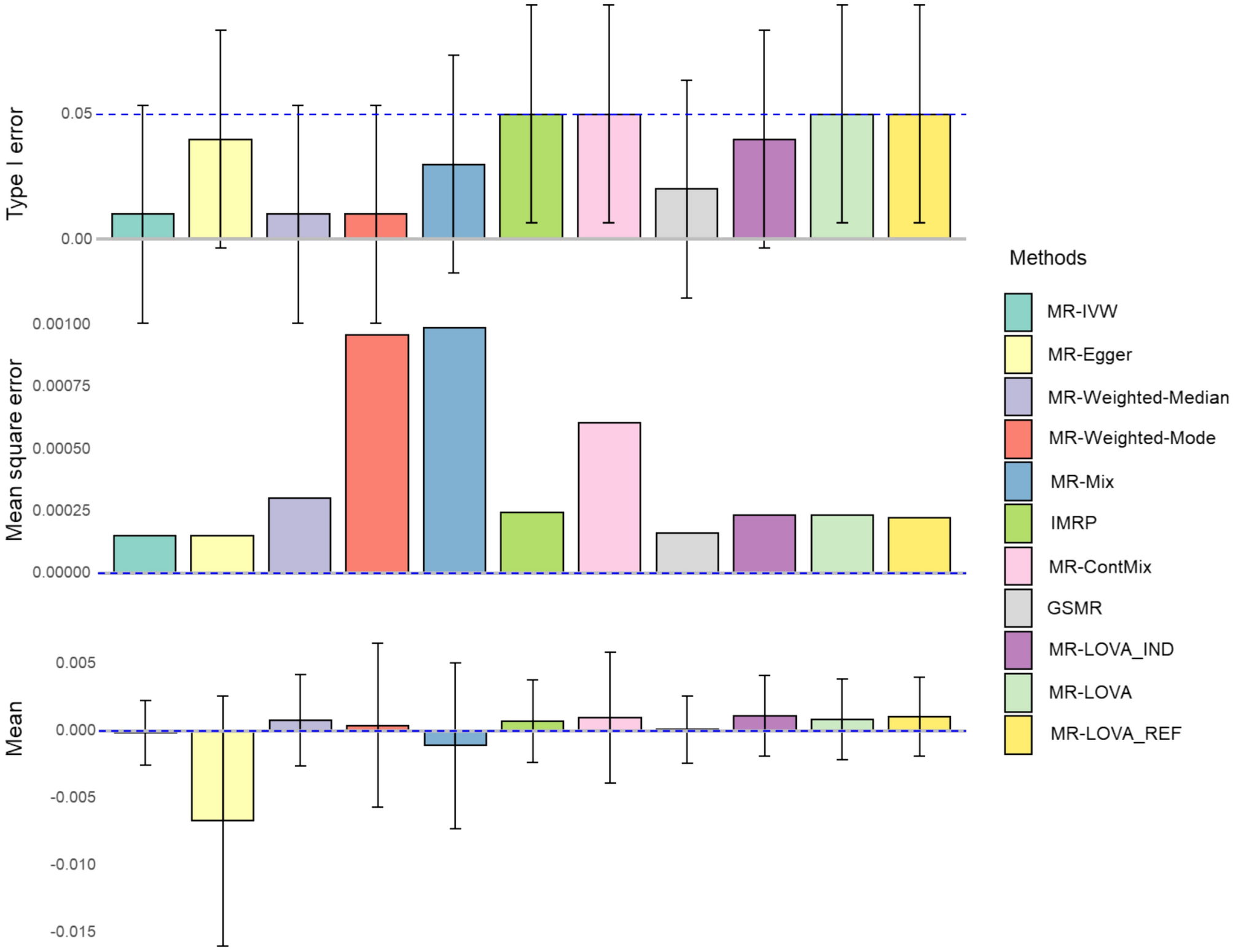
VCF files were processed, and variants were prioritized using BaseSpace Variant Interpreter® (Illumina, San Diego, CA, USA), VarSeq® (Golden Helix, Inc., Bozeman, MT, USA), Congenica (Congenica LTD, Cambridgeshire, United Kingdom) and VarSome® (Lausanne, Switzerland). All genes previously reported as causative of SRNS (Supplementary Table 1) including both exonic and intronic regions were evaluated as a first step; however, the whole genome was examined when no conclusive pathogenic/likely pathogenic variants were detected in the target genes. In the second step, we evaluated all pathogenic and likely pathogenic variants detected by the above mentioned software programs across the whole genome and not previously related to SRNS. These variants were evaluated separately in the context of patients'clinical features and through reverse phenotyping if needed and only those overlapping significantly with their corresponding patients'phenotypes were reported. Copy number variants (CNVs) and structural variants (SVs) were assessed using BAM files uploaded to VarSeq® software. The pathogenicity of suspected variants was classified according to the American College of Medical Genetics and Genomics (ACMG) (Richards et al. 2015; Tavtigian et al. 2020) and the Association for Clinical Genomic Science (ACGS) (UK 2023) guidelines. Sanger sequencing was used to confirm the variants revealed by next-generation sequencing and to confirm that novel pathogenic variants properly segregated in parents and available family members according to the reported mode of inheritance for each affected gene.
Protein structure and modeling
The 3D structure of various proteins harboring missense variants was retrieved from the Protein Data Bank https://www.rcsb.org/. If the experimental structure of a protein did not cover the specific protein domain of interest, an alternative option is to search the AlphaFold protein structure database (Jumper et al. 2021; Varadi et al. 2022). The Human Mutation Analysis (HUMA) database https://huma.rubi.ru.ac.za/ and the web server HUMA-PRIMO were used to generate a 3D structural model for mutant and wild-type proteins and an interactive homology modeling pipeline that can be used to introduce variants into protein structures (Brown and Tastan Bishop 2018). Protein structures were edited, non amino acid elements were removed, and the edited structures were saved in (.pdb) format. Structures were then visualized using the UCSF Chimera: (Pettersen et al. 2004). Mutant models were further constructed and analyzed from their wild type using the PyMOL tool: http://pymol.sourceforge.net/ (Bramucci et al. 2012). A protein stability change upon single point mutation was predicted by using MAESTRO web (Laimer et al. 2016) and I-Mutant 2.0 server (Capriotti et al. 2005). Basewise conservation was evaluated by the PhyloP100 score, which evaluates the alignment of the affected base against 100 vertebrate species. The PhyloP100 conservation scores were obtained from the University of California, Santa Cruz (UCSC) genome website (https://genome.ucsc.edu/).


Comments (0)