
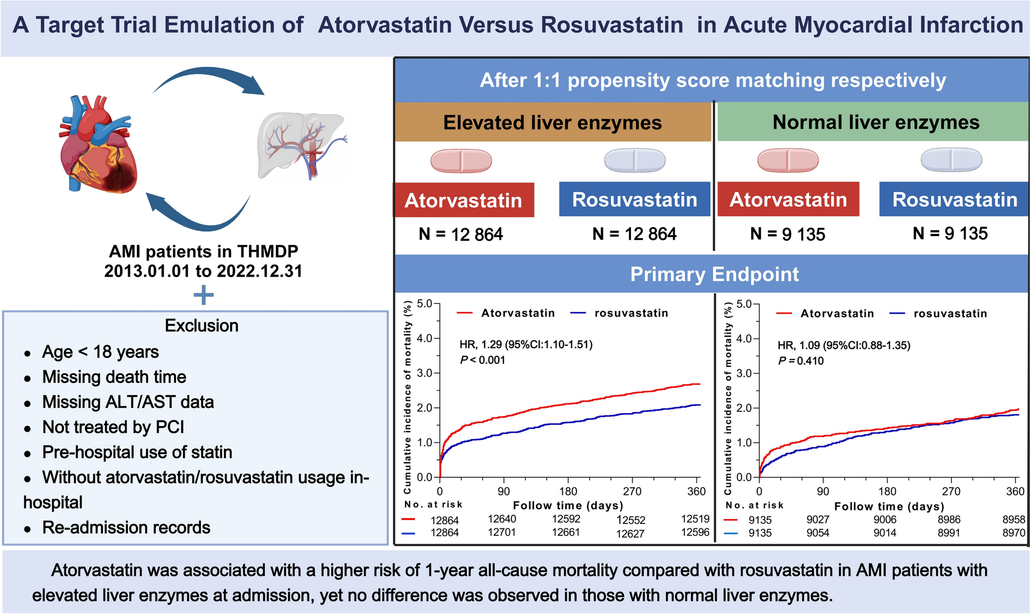
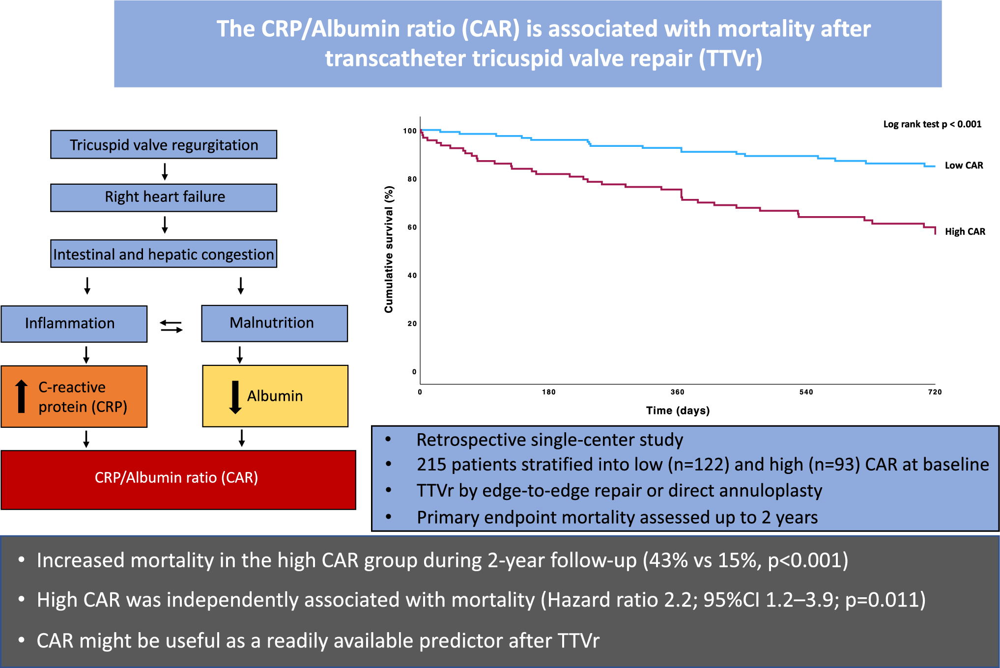
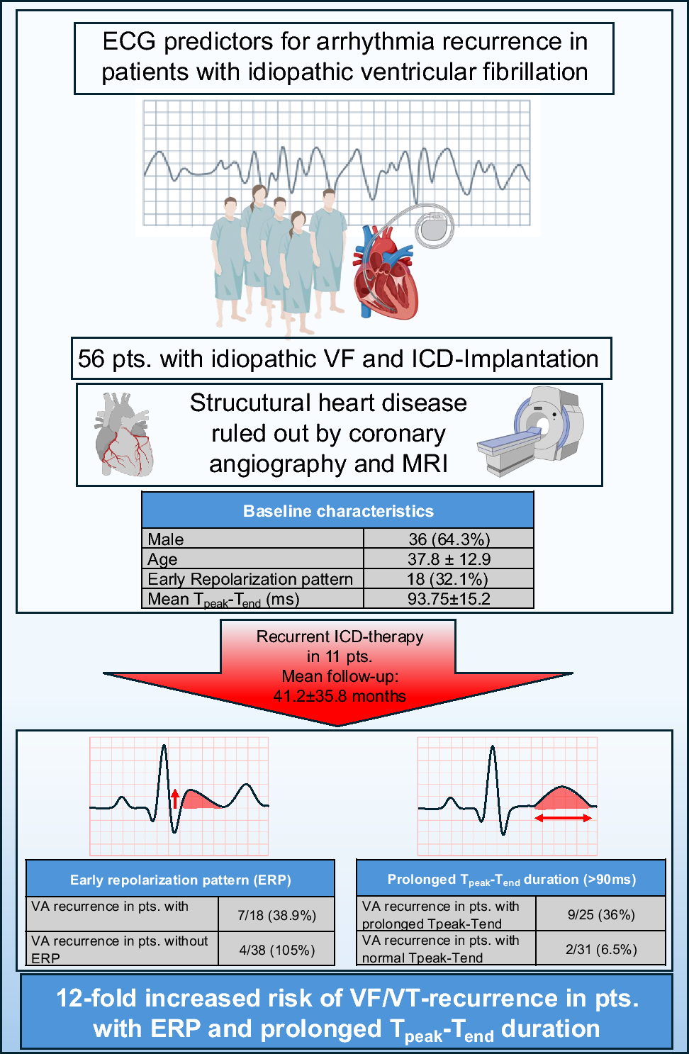
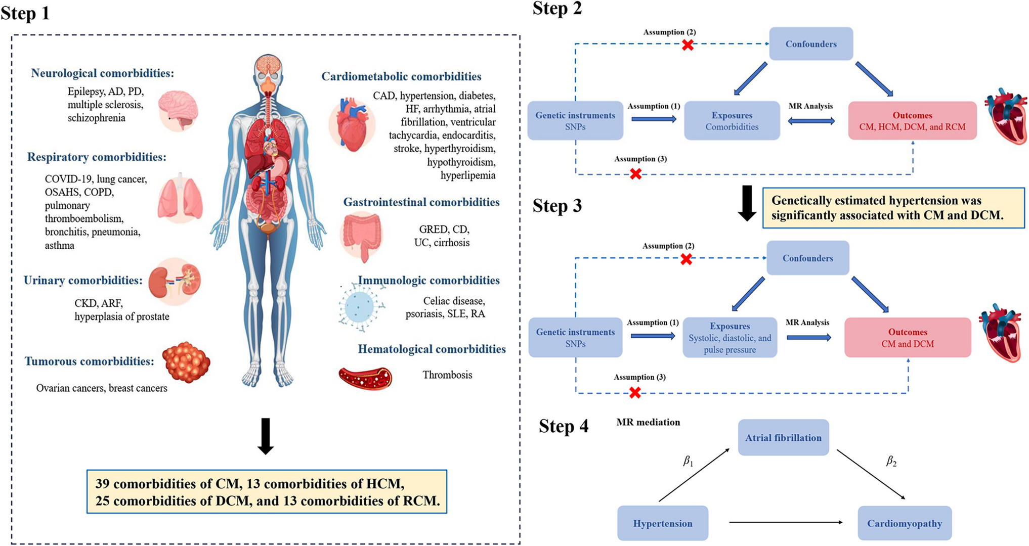
Remember me


The Stunning in Takotsubo versus Acute Myocardial Infarction study (STAMI, NCT04448639) is a prospective, observational study in which patients with TS or STEMI without preexisting cardiac dysfunction undergo echocardiography, electrocardiography, and laboratory assessment within 4 h of admission and at days 1, 2, 3, 7, 14, and 30 (Supplementary tables S1 and S2) during 2019–2024 at Sahlgrenska University Hospital, Gothenburg, Sweden. TS was defined according to the European Society of Cardiology (ESC) diagnostic criteria [9]. Patients with STEMI were required to undergo primary PCI within 6 h of symptom onset. Only women with TS or anterior STEMI were included in the analysis population. All study participants gave informed consent, and the study was approved by the Swedish Ethical Review Authority (Dnr. 2022-01003-02) and carried out in accordance with the Declaration of Helsinki. All data was handled after the EU Data Protection Directive.
EchocardiographyAll echocardiograms were performed according to a detailed echocardiographic protocol by experienced echocardiographers (Supplemental Appendix 1); each echocardiogram was analysed offline per the protocol in a blinded manner independently by two experienced echocardiographers (SJ and AP). In preparation for STAMI, we demonstrated that the intra- and inter-observer variability for the echocardiographic variables of interest is low, and the method is reliable [10]. For each of the echocardiographic variables, the mean value between the two observers were used for analyses.
DefinitionsWe defined proportion akinesia (PrA) as the proportion of the left ventricular endocardial length (as measured in the apical 2- and 4-chamber views at end-diastole) that was akinetic (Fig. 1) [10]. We defined early akinesia resolution as the difference in PrA between baseline (day 0) and day 3; and total akinesia resolution as the difference in PrA between baseline and day 30.
Fig. 1
Assessment of the proportion akinesia in the left ventricular four- (left) and two (right) chamber view. First, the left ventricular endocardial border was outlined in apical four chamber view (blue line) in end-diastole. Secondary, the endocardial border and the extent of akinesia was outlined (red line). The total extent of akinesia in millimetres in both the apical four- and two chamber views was divided by the total length in millimetres in both apical four- and two chamber views in diastole multiplied by 100. RA right atrium, RV right ventricle, LV left ventricle, LA left atrium
Primary outcomeIn this study, we defined stunning as reversible akinesia. The primary outcome was stunning resolution at 3 days, defined as early resolution of akinesia divided by total resolution of akinesia per the following formula:
$$stunning \, resolution=\frac_-_}_-_}$$
The reason for choosing resolution of akinesia rather than LVEF or LV GLS as the primary outcome is that we were primarily interested in studying the recovery of the affected myocardial segments, and remote, unaffected regions of LV can compensate for the dysfunctional myocardial segments and mitigate the reduction in both LVEF and GLS [11].
Secondary outcomesPrespecified secondary outcomes of interest were the differences between women with TS and women with STEMI over time in regards to (i) left ventricular ejection fraction (LVEF), (ii) left ventricular global longitudinal strain (GLS), (iii) wall motion score index (WMSI), (iv) tricuspid annular plane systolic excursion (TAPSE), (v) serum troponin-I, (vi) serum troponin-T, (vii) serum troponin-I/troponin-T ratio, (viii) serum NT-proBNP, (ix) serum NT-proBNP/troponin-I ratio, and (x) serum NT-proBNP/troponin-T ratio.
Statistical analysesContinuous data are reported as mean ± standard deviation for variables with normal distribution, and median (quartile 1 [Q1], quartile 3 [Q3]) for variables with skewed distribution. Categorical data are reported as frequency (percentage). Missing data in akinesia was partially imputed as follows: observations of 0% akinesia were carried forward if there was no subsequent observation of non-zero akinesia. Patients who experienced a second event during follow up were censored from the day and time of the second event. Second events included additional myocardial infarction and additional revascularisation, including staged PCI.
The primary analysis compared stunning resolution between women with TS and women with anterior STEMI. Identical tobit mixed models were fitted to model the evolution of akinesia over time. The tobit model functions as a zero-inflated and left-censored normal distribution, allowing for zero akinesia. Time was included as a fixed categorical variable, and patient-specific trajectories were modelled using “random” intercepts. TS and STEMI were fitted separately, allowing both condition-specific reference trajectories and differences in the between-patient and within-patient variation. For each condition, the stunning resolution for the median patient at day 3 was derived from the tobit mixed model using 10,000 posterior samples obtained using Markov Chain Monte Carlo (R package brms 2.21.0). The difference in stunning resolution for the median patient was obtained as the simple difference between the two posterior samples. Percentiles were used to calculate 95% Credible Intervals. Covariates were included in the model using cubic splines for age and body mass index (BMI), and as binary variables for diabetes, hypertension, and chronic obstructive pulmonary disease. The adjusted difference in stunning resolution was calculated for the median patient with median BMI, median age, and without comorbidities.
Mixed-effects linear regression was used to model the temporal variation in echocardiographic and laboratory parameters, with time as a fixed categorical effect and random intercepts assigned to each patient ID. The model was used uniformly for variables with a normal distribution (LVEF, GLS, TAPSE) and for those conforming to normality after logarithmic transformation (troponin-I, troponin-T, NT-proBNP, ratios). BMI, age, diabetes, hypertension, and chronic obstructive pulmonary disease were included as covariates in the models. All statistical analyses were performed with R version 4.4.1. Figures were designed using Affinity Designer 2 and Biorender.com.
Comments (0)