
Remember me
Non-invasive imaging methods for evaluating microstructural changes in patients with developmental dysplasia of the hip (DDH) are lacking.
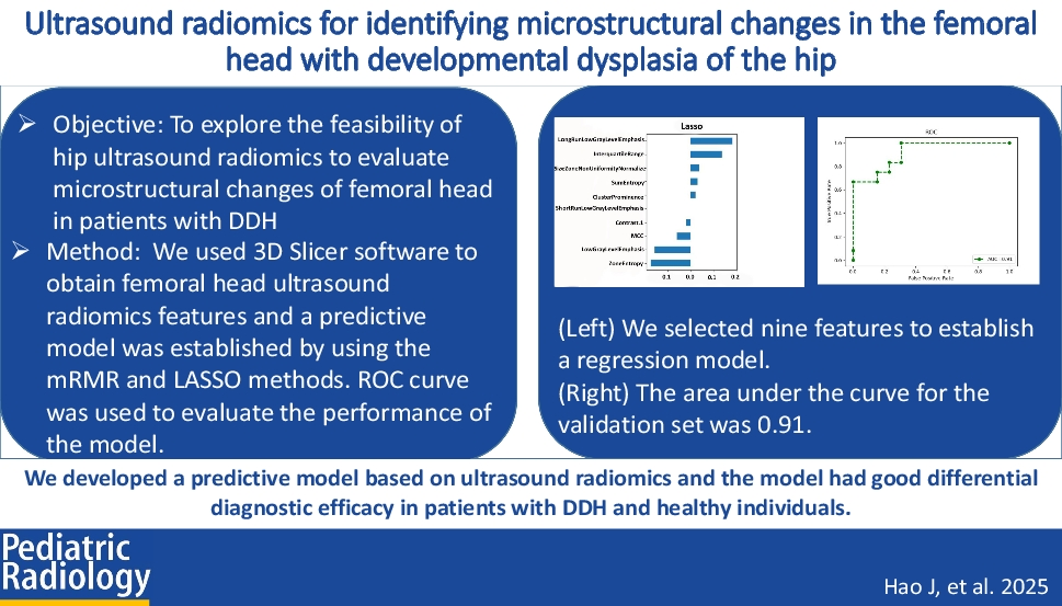
ObjectiveTo explore the feasibility of hip ultrasound radiomics to evaluate microstructural changes of the femoral head in patients with DDH.
Materials and methodsA retrospective analysis was conducted on 59 patients with DDH and 66 healthy controls who underwent hip ultrasound examination. We used three-dimensional (3D) Slicer software to obtain femoral head ultrasound radiomics features and compared them between the DDH and healthy control groups. A predictive model was established by using the maximum relevance minimum redundancy (mRMR) and least absolute shrinkage and selection operator (LASSO) methods. The receiver operating characteristic (ROC) curve was used to determine the performance of the model.
ResultsThere were significant differences (P < 0.05) in 69 ultrasound radiomics features of the femoral head between the patients with DDH and healthy controls. By using mRMR, 12 features were selected for further analysis via LASSO. We have successfully established a predictive model based on nine features. The area under the curve (AUC) of the model was 0.91 for the validation set.
ConclusionWe developed a predictive model based on ultrasound radiomics, and the model had good differential diagnostic efficacy in patients with DDH and healthy individuals.
Graphical abstract
Comments (0)