
Remember me
Osteosarcoma is the most common bone pediatric cancer, with the lung being the primary site of metastasis. A chest computed tomography scan (CT) is used to assess metastatic disease at diagnosis, classifying patients as localized or metastatic. Although there are radiological characteristics that suggest whether a lung nodule is metastatic, in daily practice, non-specific lesions on CT may complicate classification.
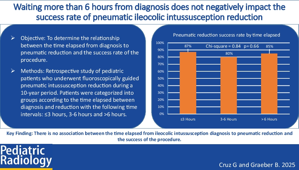
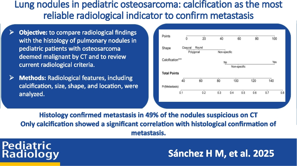
ObjectiveOur objective is to compare radiological findings with the histology of lung nodules deemed malignant by CT and to review current radiological criteria.
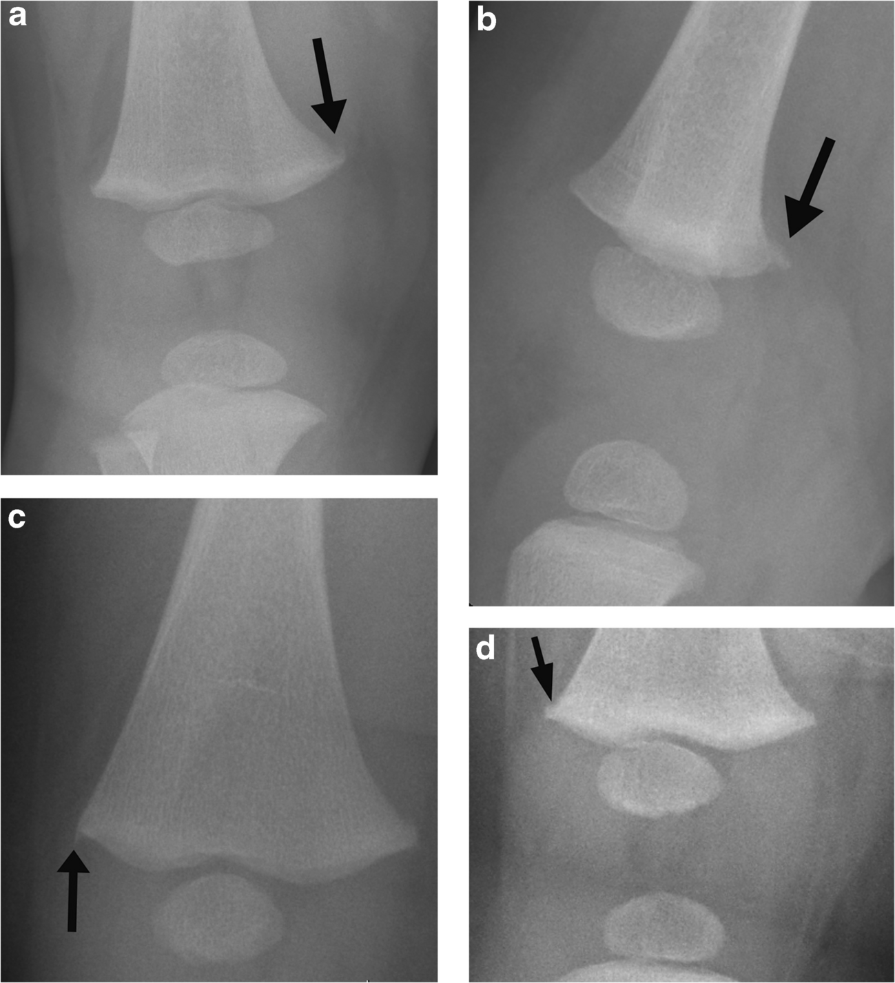
Materials and methodsA retrospective review was conducted of lung nodules in patients under 18 years old, diagnosed with osteosarcoma between 2014-2024 in a tertiary hospital. Radiological features, including calcification, size, shape, and location, were analyzed for their correlation with histological confirmation of metastasis.
ResultsIn 33 osteosarcoma patients, 116 nodules were identified as malignant by radiology. A total of 69% of patients had pulmonary nodules that met radiological criteria for malignancy during follow-up. All underwent surgical resection. Histology confirmed metastasis in 49% (57/116) of the nodules that were suspicious on CT. Only calcification showed a significant correlation with histological confirmation of metastasis.
ConclusionsCT imaging has an optimal sensitivity but low specificity for detecting lung metastases in osteosarcoma. However, we demonstrate that calcification is significantly correlated with histological confirmation of metastasis and may aid in confirming lung metastasis in osteosarcoma patients. Still, further studies are needed to refine radiological criteria to improve accuracy and reduce false positive rates.
Graphical abstract
Comments (0)