
Remember me


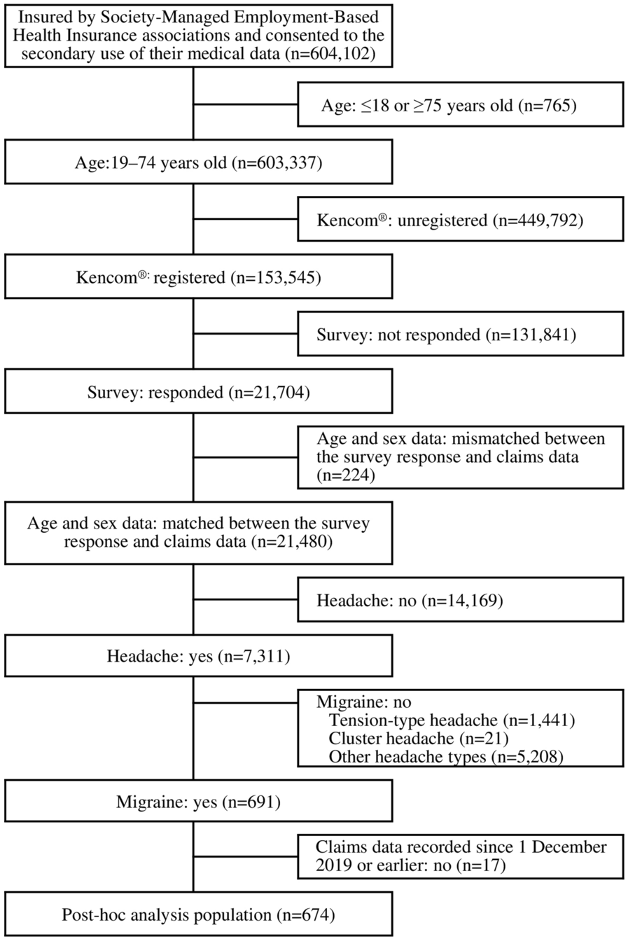

This first-in-human, Phase 1 study was conducted in 2 parts at a single center in Groningen, the Netherlands (Fig. 1). The trial enrolled healthy male and female (women of non-childbearing potential) participants aged between 18 and 55 years, body mass index of 18–30 kg/m2 , and with normal vital signs and laboratory parameters at baseline.
Fig. 1
Study design. †Preliminary safety and PK data from the first 3 dose groups of SAD was used to initiate the dose 1 in the MAD study. CFE prototype formulation of eclitasertib, DE dose escalation, DS drug substance in capsule form, eclit eclitasertib, MAD multiple ascending dose, pbo placebo, QD once daily, R randomization, SAD single ascending dose, WO washout
Part 1 consisted of 2 subparts. Part 1a was a double-blind, randomized, placebo-controlled, sequential single oral ascending dose (SAD) study with 6 cohorts of 8 participants each randomized 3:1 to receive single dose of eclitasertib (10 mg, 30 mg, 100 mg, 200 mg, 400 mg, or 800 mg) or placebo to evaluate the safety, PK, and PD of a single dose of eclitasertib. The six sequential ascending cohorts were administered a single dose of eclitasertib orally in the morning in fasted conditions (for at least 10 h). The first cohort (10 mg eclitasertib) consisted of 2 sentinel participants who were dosed on the first day, at least 15 min apart. Part 1b was an open-label, randomized, three-sequence, cross-over design study to evaluate the relative bioavailability between drug substance (DS) in capsule form used in the SAD and multiple ascending dose study (MAD) parts and prototype formulation for Phase 2 studies [capsule formulation, with drug substance formulated along with excipients and a granulation step (CFE)] and the effect of food on PK of CFE in an independent cohort of 10 participants,. Study participants received either a single dose of 100 mg DS under fasted condition, a single dose of 100 mg CFE under fasted condition, or a single dose of 100 mg CFE after a high-fat meal; the order in which they received treatments was determined by the assigned sequence during randomization.
Part 2 was a double-blind, randomized, placebo-controlled, sequential MAD with 4 cohorts of up to 10 participants each randomized 4:1 to receive eclitasertib (50 mg, 100 mg, 200 mg, or 600 mg once daily) or placebo orally for 14 days to evaluate the safety, PK, and PD of multiple doses of eclitasertib. Emerging safety data and available PK data from preceding cohorts were reviewed at dose escalation meetings prior to dose escalation.
The study protocol was approved by independent ethics committee [The Medical Research Ethics Committee of the ‘Stichting Beoordeling Ethiek Biomedisch Onderzoek’ (Foundation of Ethics in Biomedical Research), Assen, The Netherlands] and/or institutional review boards, and was conducted in accordance with the Declaration of Helsinki and the International Council for Harmonization guidelines for Good Clinical Practice.
The participants were informed of the risks and benefits of the trial, and that they could withdraw from the trial at any time for any reason. Written informed consent was obtained from all the participants before any study-related interventions were administered.
Study EndpointsAssessment of safety of eclitasertib was the primary endpoint. Secondary endpoint included evaluation of plasma PK parameters (Part 1 and Part 2). The exploratory endpoint was the reduction in RIPK1 phosphorylation at serine 166 (pS166-RIPK1) in human peripheral blood mononuclear cells (PBMC), measured after a single dose (Part 1) or 14 days of eclitasertib treatment (Part 2).
Safety AssessmentsAssessment of safety and tolerability included incidence of AEs and their severity, AEs of special interest [AESI; new onset acute lymphadenopathy, anemia, elevated alanine aminotransferase (ALT) levels], abnormal clinical laboratory findings, vital signs, electrocardiogram (ECG) parameters, and physical examinations in parts 1 and 2. AEs that could be attributed to invasive procedures for sample collections for PK, PD, and laboratory analyses were classified as ‘medical device-site reactions’ and included AE terms medical device-site reaction, vessel puncture-site hematoma/pain, catheter-site pain/hematoma, and catheter-site-related reactions.
Pharmacokinetic AssessmentsPlasma and urine concentrations of eclitasertib were analyzed using validated liquid chromatography coupled with tandem mass spectrometry (LC–MS/MS) with a lower limit of quantification of 0.2 ng/mL.
Plasma concentrations were analyzed using two validated and cross-validated LC–MS/MS methods with two different calibration ranges (low range 0.2–200 ng/mL; high range 20–20,000 ng/mL). The low calibration range assay method was used to assay all predose samples of all cohorts and all samples from the doses 10–200 mg in Part 1a and 50 mg in Part 2, and to assay samples collected around Tmax of placebo participants. The high calibration assay was used to evaluate the samples of the last cohorts (from 400 mg in Part 1a and from 100 mg Part 2). However, for time points in the elimination phase when the concentrations were below the limit of quantification (20 ng/mL), the samples were re-assayed with the low calibration range assay [lower limit of quantification (LLOQ) of 0.2 ng/mL]. Plasma samples were collected at pre-dose (0 h) and at 30 min, 1 h, 2 h, 3 h, 4 h, 6 h, 8 h, 10 h, 12 h, 16 h, 24 h, 48 h and 72 h post-dose in the SAD part, and at pre-dose (0 h), at 30 min, 1 h, 2 h, 3 h, 4 h, 6 h, 8 h, 10 h, 12 h, 16 h post-dose on day (D) 1 and D14, pre-dose on D2, D3, D4, D5, D7, D10, D13, and 24 h, 48 h and 72 h post-dose on D14 in the MAD part. Urine samples were collected soon after eclitasertib administration (0–4 h), 4–12 h, and 12–24 h post-dose in the SAD part and after the last administration of eclitasertib on D14 in the MAD part.
PK parameters [maximum plasma concentration (Cmax), time to maximum concentration (Tmax), area under curve (AUC0–24 h, area under the plasma concentration versus time curve from 0 to 24 h) and (AUC, area under the plasma concentration versus time curve extrapolated to infinity), terminal half-life (t1/2z), apparent total body clearance (CL/F)] were determined by non-compartmental analysis (NCA) using PKDMS Version 3.1 using Phoenix 8.1 (Certara™).
4-β-hydroxycholesterol was assessed using an ultra-high-performance liquid chromatography with tandem mass spectrometric method with a calibration range of 5–150 ng/mL.
Pharmacodynamic AssessmentsRIPK1 undergoes autophosphorylation at S166 upon activation. Human PBMCs express detectable endogenous RIPK1 [13, 14]. Therefore, the ability of eclitasertib to inhibit RIPK1 activity, as measured by pS166-RIPK1 levels in PBMC lysates isolated from blood samples, was used as a PD marker to assess the peripheral target engagement [13, 14]. Levels of S166 phosphorylated RIPK1 in unstimulated PBMCs were assessed as described previously, but without TSZ (TNFα, SM-164, Z-VAD-FMK) stimulation [13,14,15]. In brief, isolated PBMCs were resuspended in autologous plasma and incubated for 150 min at 37 °C, and 5% CO2. PBMCs were lysed in 1X CST buffer (Cell Signaling Technologies, Leiden, the Netherlands) and subsequently centrifuged, and the supernatant was frozen at − 80 °C until assayed [13]. pS166-RIPK1 was detected with a meso scale discovery (MSD) platform-based immunoassay. Each well of a streptavidin small spot 96-well plate coated with biotinylated mouse anti-RIPK1 antibody was incubated with PBMC lysate for 2 h followed by rabbit anti-pS166 RIPK1 for 1 h. pS166-RIPK1 levels were detected with SULFO tagged goat anti-rabbit secondary antibody using a using a Sector S600 plate reader (MSD) [13]. Samples for the measurement of pS166-RIPK1 levels in human PBMC lysates were collected at pre-dose (0 h), and at 1 h, 6 h, 12 h, 24 h, 48 h, and 72 h post-dose in the SAD part and at pre-dose (0 h on D1 and D14), and at 2 h, 6 h, and 12 h post-dose on D14, and on D15, D16, and D17.
Statistical AnalysisSafety, PK, and PD data were summarized with descriptive statistics using SAS version 9.4 (SAS Institute, NC, USA).
Dose proportionality was assessed using an empirical power model (pharmacokinetic parameter = α × doseβ), along with an “estimation” interpretation, as described by Gough et al. [16]. The power model was fit on the log-transformed scale using SAS version 9.4: log (parameter) = log (α) + β × log (dose) as described previously [13, 15].
To estimate the effect of food on PK parameters, Cmax, and AUC, the difference between the fasted and fed conditions (including data on participants receiving CFE only) was assessed on log-transformed parameters with a linear model,
$$}\left( }} \right) \, = } + } + } + }$$
with a fixed term for sequence, period, and food condition, and with an unstructured 2 × 2 matrix of food condition variances and covariance for subject within sequence.
To estimate bioavailability differences between DS and CFE formulation in the fasted state, Cmax, and AUC were assessed on log-transformed parameters with a linear model,
$$}\left( }} \right) \, = } + } + } + }$$
with a fixed term for sequence, period, formulation DS, and formulation CFE, and with an unstructured 2 × 2 matrix of formulation variances and covariance for subject within sequence.
The reduction in RIPK1 S166 phosphorylation in human PBMC lysates was analyzed as raw values and absolute and percent change from baseline. Results were summarized by cohort using descriptive statistics.
Comments (0)